Molecular Imaging

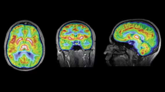
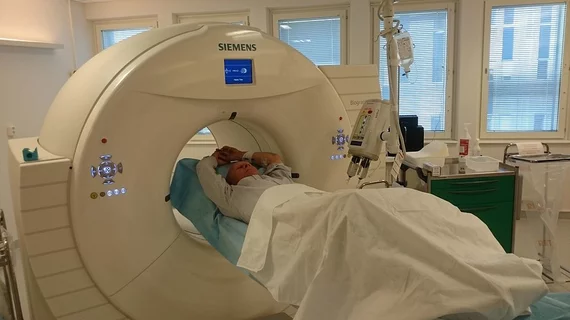
Molecular imaging (also called nuclear medicine or nuclear imaging) can image the function of cells inside the body at the molecular level. This includes the imaging modalities of positron emission computed tomography (PET) and single photon emission computed tomography (SPECT) imaging. How does PET and SPECT imaging work? Small amounts of radioactive material (radiopharmaceuticals) injected into a patient. These can use sugars or chemical traits to bond to specific cells. The radioactive material is taken up by cells that consume the sugars. The radiation emitted from inside the body is detected by photon detectors outside the body. Computers take the data to assemble images of the radiation emissions. Nuclear images may appear fuzzy or ghostly rather than the sharper resolution from MRI and CT.  But, it provides metabolic information at a cellular level, showing if there are defects in the function of the heart, areas of very high metabolic activity associated with cancer cells, or areas of inflammation, data not available from other modalities. These noninvasive imaging exams are used to diagnose cancer, heart disease, Alzheimer’s and Parkinson’s disease, bone disorders and other disorders. 

PET measure sheds light on response to chemo for breast cancer

The change in total lesion glycolysis measured prior to and after two cycles of chemotherapy provided a better prediction of response to chemotherapy than other PET measures, including change in standardized uptake value, according to a study published online Jan. 17 in The Journal of Nuclear Medicine.

The Balancing Act PET/CT: Ensuring Image Quality, Controlling Dose

New methods for PET/CT imaging are being developed for imaging cancer patients without overexposing them to radiation while also guaranteeing excellent image quality.

SPECT/CT’s Role in Post-Transplant Infection Imaging

Timely and accurate diagnosis and treatment of infection in post-transplant organ patients is critical to patient survival and the prevention of organ rejection. 

Nuclear Cardiology’s Next Step

 Nuclear cardiology is on the dawn of a new technology—IQ•SPECT promises to reduce acquisition time, add hybrid or fusion imaging with CT and maximize efficiency in SPECT/CT scanning.

NOPR: A Landmark Study

Initial Results from National Oncologic PET Registry (NOPR) have been published recently in the Journal of Clinical Oncology. The results provide impressive evidence for the substantial impact of PET imaging across all cancers.

Evidence-based Medicine Points to Wider Role for Molecular Imaging in Patient Care

Evidence-based medicine has been embraced as a practice to increase healthcare expenditures and as a tool to improve patient care, ensuring that patients receive the best resources for their care, without unnecessary and wasteful procedures. Clinical results are proving molecular imaging’s merits in a wider number of applications. 

Changing Treatment Early Enough to Matter

This issue we are discussing several important topics in molecular imaging and medicine. One addresses the need for developing tools not only to monitor therapeutic responses, but also to stratify patients into the treatments that are most likely to result in significant responses. This concept can be applied not only to oncology, but also is highly relevant in patients with neurologic and cardiac diseases.

Assessing Cancer Therapy & Infection

This issue features several topics of critical importance in the field of imaging.