Molecular Imaging

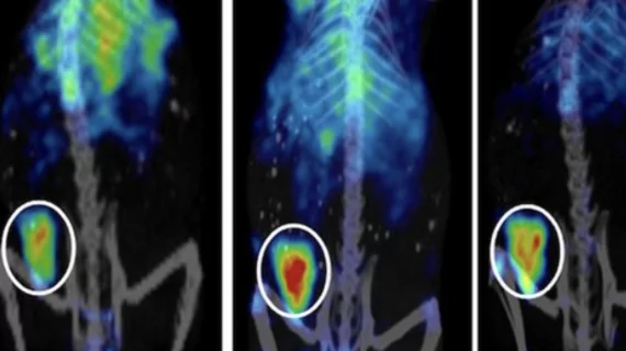
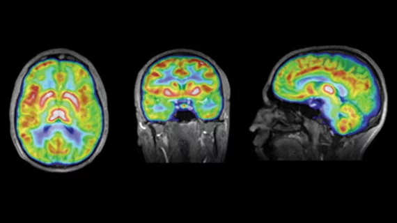
Molecular imaging (also called nuclear medicine or nuclear imaging) can image the function of cells inside the body at the molecular level. This includes the imaging modalities of positron emission computed tomography (PET) and single photon emission computed tomography (SPECT) imaging. How does PET and SPECT imaging work? Small amounts of radioactive material (radiopharmaceuticals) injected into a patient. These can use sugars or chemical traits to bond to specific cells. The radioactive material is taken up by cells that consume the sugars. The radiation emitted from inside the body is detected by photon detectors outside the body. Computers take the data to assemble images of the radiation emissions. Nuclear images may appear fuzzy or ghostly rather than the sharper resolution from MRI and CT.  But, it provides metabolic information at a cellular level, showing if there are defects in the function of the heart, areas of very high metabolic activity associated with cancer cells, or areas of inflammation, data not available from other modalities. These noninvasive imaging exams are used to diagnose cancer, heart disease, Alzheimer’s and Parkinson’s disease, bone disorders and other disorders. 

PET & PET/CT Earn Role in Oncologic Therapeutic Response

Slowly but steadily, radiation oncologists are adopting PET and PET/CT to measure the early response of cancers to radiotherapy and other treatments. And progress has been significant.

Treatment Monitoring with FDG PET/(CT)

The cover story in this issue discusses the role of PET imaging for assessing therapeutic responses in cancer. Over the past 15 years, a large body of evidence has demonstrated that FDG-PET imaging is uniquely useful to assess treatment responses in cancer patients.

Nuclear Imaging Helps Manage Heart Failure Patients

Using both PET and SPECT may have the potential to provide clinically useful data to enable better stratification and favorable treatments for heart failure patients.

Down Deep Differentiating Sentinel Nodes

Accurate localization of sentinel nodes with SPECT/CT has expanded beyond breast cancer and melanoma to head and neck cancer.

Offering Sharper Tools for Neurological Disorders

SPECT and PET-based strategies have started to refine diagnosis and treatment planning for Parkinsons disease and epilepsy.

Preclinical Study Digest

Imaging blood perfusion in pig livers; A molecular theranostics primer; Mice provide a biomarker roadmap for human approach; Simultaneous PET/3D FOT is feasible

ASNC Commits to Patient-Centered Care

The 16th annual scientific session of the American Society of Nuclear Cardiology (ASNC), Sept. 8-11, brings together scientists, clinicians and industry to establish and discuss optimal standards of imaging care.

AJR: A molecular theranostics primer

Molecular theranostics holds promise, offered a review published in the August issue of American Journal of Roentgenology.