Molecular Imaging

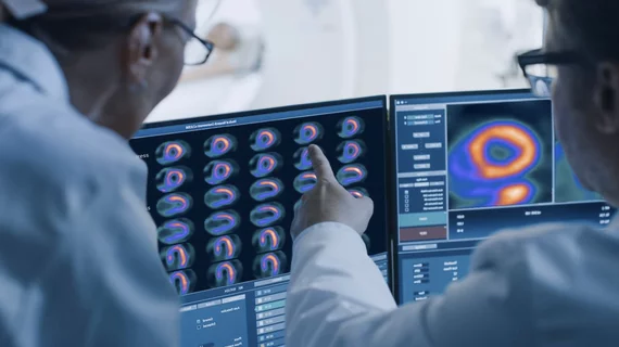
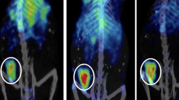
Molecular imaging (also called nuclear medicine or nuclear imaging) can image the function of cells inside the body at the molecular level. This includes the imaging modalities of positron emission computed tomography (PET) and single photon emission computed tomography (SPECT) imaging. How does PET and SPECT imaging work? Small amounts of radioactive material (radiopharmaceuticals) injected into a patient. These can use sugars or chemical traits to bond to specific cells. The radioactive material is taken up by cells that consume the sugars. The radiation emitted from inside the body is detected by photon detectors outside the body. Computers take the data to assemble images of the radiation emissions. Nuclear images may appear fuzzy or ghostly rather than the sharper resolution from MRI and CT.  But, it provides metabolic information at a cellular level, showing if there are defects in the function of the heart, areas of very high metabolic activity associated with cancer cells, or areas of inflammation, data not available from other modalities. These noninvasive imaging exams are used to diagnose cancer, heart disease, Alzheimer’s and Parkinson’s disease, bone disorders and other disorders. 

Positron subsidiary nets FDA drug master file for Sr-82

Positron's subsidiary, Manhattan Isotope Technology, has received an acknowledgment letter and Drug Master File number assignment from the FDA for its strontium-82 drug substance.

Gamma imaging proves its prowess in DCIS detection

Breast-specific y camera scintigraphy is a highly specific tool for ductal carcinoma in situ (DCIS) detection, and provides 100 percent sensitivity when paired with mammography, according to a study published in the October issue of the Journal of Nuclear Medicine.

Calif. governor signs breast density notification bill

California Gov. Jerry Brown has signed a bill requiring physicians to give extra warnings to women with dense breast tissue, informing the women that additional screening through ultrasound or MRI, as opposed to mammography alone, may be recommended.

Racial, socioeconomic disparities pervade PET imaging

Use of PET among Medicare beneficiaries with cancer increased from 2004 to 2008, but the increase was not even between sociodemographic groups as the modality had higher utilization among whites and those in higher income zip codes, according to a study published in the September issue of Journal of the American College of Radiology.

Perceived Radiation Risks, Alzheimers Disease and Next-generation Imaging Approaches

This issue provides discussions about potential risks associated with diagnostic tests in pediatric patients, PET probes for imaging neuroinflammation and neurodegenerative diseases, as well as emerging hybrid imaging technologies such as PET/MRI.

Balancing Risk-benefit in Pediatric Nuclear Medicine Far from Childs Play

Professional societies have stepped up efforts to minimize administered activity, based on updated dosage cards and guidelines. Medical physicists recognize that while these protocols are helpful, there is room for improvement.

I-131 Best Practice Or In Need of Use Revision?

Iodine-131 is lauded for its specificity in targeting thyroid lesions. Yet, some experts are debating whether the treatment should be used across the board or stratified.

Targeting Lesion Localization: A New Era

While whole-body oncology imaging advances are still emerging, the best modality remains to be seen.