Molecular Imaging

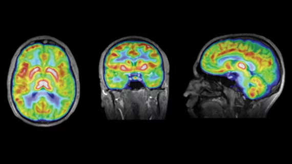
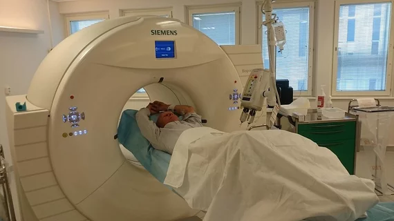
Molecular imaging (also called nuclear medicine or nuclear imaging) can image the function of cells inside the body at the molecular level. This includes the imaging modalities of positron emission computed tomography (PET) and single photon emission computed tomography (SPECT) imaging. How does PET and SPECT imaging work? Small amounts of radioactive material (radiopharmaceuticals) injected into a patient. These can use sugars or chemical traits to bond to specific cells. The radioactive material is taken up by cells that consume the sugars. The radiation emitted from inside the body is detected by photon detectors outside the body. Computers take the data to assemble images of the radiation emissions. Nuclear images may appear fuzzy or ghostly rather than the sharper resolution from MRI and CT.  But, it provides metabolic information at a cellular level, showing if there are defects in the function of the heart, areas of very high metabolic activity associated with cancer cells, or areas of inflammation, data not available from other modalities. These noninvasive imaging exams are used to diagnose cancer, heart disease, Alzheimer’s and Parkinson’s disease, bone disorders and other disorders. 

Biomarker may measure brain tumor response to RT

Diffusion abnormality index has emerged as a potential biomarker to measure brain tumor response to radiation therapy (RT), according to research presented at the 2013 Cancer Imaging and Radiation Therapy Symposium Feb. 9 in Orlando, Fla. The symposium is sponsored by the American Society for Radiation Oncology and the Radiological Society of North America.

Speedy in-room PET shows potential for planning proton therapy

In-room PET scanning during proton therapy is feasible for monitoring treatment ranges and helps overcome logistical issues inherent with transporting a patient to a PET scanner outside the treatment room, according to a small clinical trial published online Feb. 5 in the International Journal of Radiation Oncology*Biology*Physics.

SUVmax indicates progression-free survival in Stage 1 lung cancer

SUVmax (maximum standardized uptake value) may provide a marker to indicate progression-free survival in Stage 1 nonsmall cell lung cancer patients treated with radiation therapy, according to research presented Feb. 9 at the 2013 Cancer Imaging and Radiation Therapy Symposium in Orlando, Fla.

CZT SPECT accurately quantifies ejection fraction

While myocardial perfusion imaging using cadmium-zinc-telluride (CZT) SPECT cameras accurately measures left ventricular ejection fraction, the method underestimates left ventricular volumes, according to a study published online Feb. 5 in the Journal of Nuclear Medicine.

Dual-time-point PET/CT may help determine malignancy in kids

Semiquantitative analysis of dual-time-point FDG PET data increased specificity for pediatric malignancies compared with single-time-point PET imaging, according to a study published in the February issue of American Journal of Roentgenology.

Flurpiridaz F-18 PET superior to SPECT

Flurpiridaz F-18 PET myocardial perfusion imaging (MPI) outperformed SPECT MPI in terms of image quality, diagnostic certainty and sensitivity, according to a study published Jan. 22 in the Journal of the American College of Cardiology.

18F-ISO-1 shows promise for imaging cell proliferation

The first human study of the cell proliferation marker F18-ISO-1 indicated its safety and feasibility and suggested it may be used to stratify patients into high and low Ki-67 proliferative groups. The study was published online Jan. 28 in The Journal of Nuclear Medicine.

Cardiac CT does not stack up to MPI for ischemia detection

Resting cardiac CT performed on a 64-slice system does not reliably detect myocardial ischemia compared with radionuclide myocardial perfusion imaging (MPI), according to a study published in the February issue of the American Journal of Roentgenology.