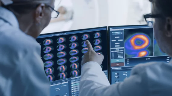
Molecular Imaging

Molecular imaging (also called nuclear medicine or nuclear imaging) can image the function of cells inside the body at the molecular level. This includes the imaging modalities of positron emission computed tomography (PET) and single photon emission computed tomography (SPECT) imaging. How does PET and SPECT imaging work? Small amounts of radioactive material (radiopharmaceuticals) injected into a patient. These can use sugars or chemical traits to bond to specific cells. The radioactive material is taken up by cells that consume the sugars. The radiation emitted from inside the body is detected by photon detectors outside the body. Computers take the data to assemble images of the radiation emissions. Nuclear images may appear fuzzy or ghostly rather than the sharper resolution from MRI and CT.  But, it provides metabolic information at a cellular level, showing if there are defects in the function of the heart, areas of very high metabolic activity associated with cancer cells, or areas of inflammation, data not available from other modalities. These noninvasive imaging exams are used to diagnose cancer, heart disease, Alzheimer’s and Parkinson’s disease, bone disorders and other disorders. 

Phase III Alzheimer’s treatment study misses the mark

Baxter announced results of its Phase III clinical study of immunoglobulin (IG). In the randomized, double-blind, placebo-controlled, multi-center trial, IG did not meet its co-primary endpoints of reducing cognitive decline and preserving functional abilities in patients with mild to moderate Alzheimer's disease after 18 months of treatment. Given the findings, Baxter said it will re-evaluate its approach for its Alzheimer’s program.

The power of exercise: SPECT MPI in active patients may be unnecessary

Many older patients undergoing an exercise stress test may not need molecular imaging to provide a clear picture of cardiovascular health. Evidence suggests that SPECT MPI may not provide any additional information for those who are nimble enough to reach a certain level of exertion and show no signs of CAD or cardiovascular distress. These and similar findings point to potential radioisotope injection guidelines to reduce radiation dose and cost of healthcare for these patients, according to research presented at the International Conference on Nuclear Cardiology and Cardiac CT, held May 5 to 8 in Berlin.

MILabs Introduces New Benchtop Product Line

MILabs, B.V. the Netherlands‐based innovator and manufacturer of pre‐clinical SPECT/PET/CT imaging devices used in biomedical research, today announces the release of the new mini product family ‐ an easy‐to‐use series of benchtop SPECT and PET imagers for small animals that combine simplified workflow, compact dimensions and affordable pricing to bring high‐performance SPECT/PET within everyone’s reach.

Spotlight: Breast-specific gamma imaging finds more cancer than mammo

Molecular breast imaging, also known as breast-specific gamma imaging (BSGI), was found to be significantly more sensitive compared to mammography. In fact, BSGI found lesions in 4 percent of women who received a clean bill with mammography, according to a meta-analysis published online by the European Journal of Nuclear Medicine and Molecular Imaging.

Not So Fast: Signs of Lewy body dementia not seen by dopamine SPECT

For patients suspected to have dementia with Lewy bodies (DLB), SPECT brain imaging of the dopamine transporter (DAT) system has been considered a possible means of evaluating classic symptoms of the disease—hallucinations, cognitive fluctuations, dementia and parkinsonism—and disease-specific changes in striatal dopaminergic function, but researchers have found no such positive connection, according to a study published May 1 by the Journal of Nuclear Medicine.

PERCIST’s Potential: PET & Quantification

The PET Response Criteria in Solid Tumors (PERCIST) aim to propel PET from the murky realm of qualitative imaging to a quantitative breakdown of response to cancer treatment.

SNMMI Treks North to Vancouver

The Society of Nuclear Medicine and Molecular Imaging (SNMMI) heads north June 8 to June 12 to Vancouver for its annual meeting, dubbed as the premiere educational and networking event in molecular imaging and nuclear medicine.

Thumbnail

FDG PET/CT points out malignant lymph nodes inside the breast

Poor patient outcomes are associated with primary breast cancer that has metastasized to internal mammary lymph nodes. Researchers compared F-18 FDG PET/CT with results of cytopathologic testing from image-guided fine-needle aspiration, revealing a high percentage of proven metastases from positive FDG PET scans, according to a study published in the May issue of the American Journal of Roentgenology.