Molecular Imaging

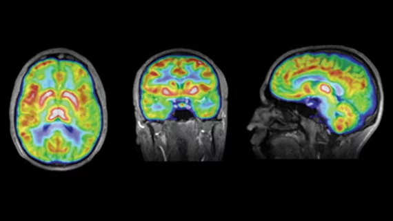
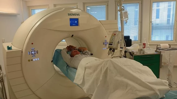
Molecular imaging (also called nuclear medicine or nuclear imaging) can image the function of cells inside the body at the molecular level. This includes the imaging modalities of positron emission computed tomography (PET) and single photon emission computed tomography (SPECT) imaging. How does PET and SPECT imaging work? Small amounts of radioactive material (radiopharmaceuticals) injected into a patient. These can use sugars or chemical traits to bond to specific cells. The radioactive material is taken up by cells that consume the sugars. The radiation emitted from inside the body is detected by photon detectors outside the body. Computers take the data to assemble images of the radiation emissions. Nuclear images may appear fuzzy or ghostly rather than the sharper resolution from MRI and CT.  But, it provides metabolic information at a cellular level, showing if there are defects in the function of the heart, areas of very high metabolic activity associated with cancer cells, or areas of inflammation, data not available from other modalities. These noninvasive imaging exams are used to diagnose cancer, heart disease, Alzheimer’s and Parkinson’s disease, bone disorders and other disorders. 

Algorithm delivers quantitative F-18 FDG PET partial volume correction

The use of algorithms is changing the game for close-as-possible assessment of tumor volume and resolution recovery from PET cancer imaging, especially small objects affected by partial volume effects. Researchers conducting phantom studies have optimized quantitative tumor delineation of F-18 FDG PET imaging with two specialized algorithms that provide accurate partial volume correction for volumes as small as one-third of a milliliter, according to research published May 8 in the Journal of Nuclear Medicine.

Can Molecular Imaging Usher in Personalized Medicine?

I have been struggling recently with the notion that genomics is going to revolutionize our ability to diagnose and treat disease. Why? A basic tenet of information theory is simple: the more precisely you can measure something, the less information it contains.

Storming the Gates: Sentinel Lymph Node Targeting & Assessment

Intraoperative lymphatic mapping provides surgeons, oncologists and referring physicians with vital information about potential malignancy in the lymphatic system, especially that of sentinel lymph nodes—usually the first check point for the diasporas of metastatic cancer cells that drain from primary tumors. Patients have a better chance of avoiding the increased morbidity associated with extensive nodal dissection by undergoing a biopsy of the sentinel lymph nodes most likely to contain metastatic disease.

Targeted Radioisotope Therapy Extends Life for Prostate Cancer Patients

Prostate cancer patients with advanced tumors that have spread to bone have a poor prognosis; men with castrate-resistant prostate cancer generally live three to five years after diagnosis.

Imaging Parkinson’s: The Search for Biomarkers

The days of the clinical exam leading the way in evaluating Parkinson’s disease are on the way out. A better understanding of how to use imaging has led to advances in diagnosing and monitoring the condition, and may hold the key to evaluating effective treatment.

PERCIST: PET Interpretation Improved

The PET Response Criteria in Solid Tumors (PERCIST), published in May 2009 in the Journal of Nuclear Medicine, were developed with a lofty goal: to shift PET from the qualitative imaging realm to a quantitative assessment of response to cancer treatment.

In Translation: Easing PET/MR into Clinical Practice

PET/MR is slowly carving out a space with an increasing number of major institutions across the globe taking advantage of the hybrid system for a widening range of research applications.

F-18 FLT PET highlights cancer proliferation

In a review of available PET imaging biomarkers that focus on cellular proliferation as an important key to cancer detection and therapy monitoring, F-18 FLT stood out as a game-changer for its ability to hone in on processes specific to cancer growth. However, in many studies, tracer uptake was underwhelming when compared to standard FDG, according to a scientific paper published May 14 in the Journal of Nuclear Medicine.