Molecular Imaging

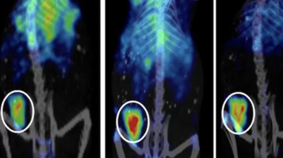
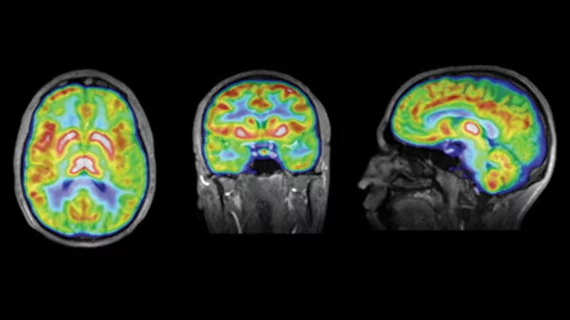
Molecular imaging (also called nuclear medicine or nuclear imaging) can image the function of cells inside the body at the molecular level. This includes the imaging modalities of positron emission computed tomography (PET) and single photon emission computed tomography (SPECT) imaging. How does PET and SPECT imaging work? Small amounts of radioactive material (radiopharmaceuticals) injected into a patient. These can use sugars or chemical traits to bond to specific cells. The radioactive material is taken up by cells that consume the sugars. The radiation emitted from inside the body is detected by photon detectors outside the body. Computers take the data to assemble images of the radiation emissions. Nuclear images may appear fuzzy or ghostly rather than the sharper resolution from MRI and CT.  But, it provides metabolic information at a cellular level, showing if there are defects in the function of the heart, areas of very high metabolic activity associated with cancer cells, or areas of inflammation, data not available from other modalities. These noninvasive imaging exams are used to diagnose cancer, heart disease, Alzheimer’s and Parkinson’s disease, bone disorders and other disorders. 

Targeted Radioisotope Therapy Extends Life for Prostate Cancer Patients

Prostate cancer patients with advanced tumors that have spread to bone have a poor prognosis; men with castrate-resistant prostate cancer generally live three to five years after diagnosis.

Imaging Parkinson’s: The Search for Biomarkers

The days of the clinical exam leading the way in evaluating Parkinson’s disease are on the way out. A better understanding of how to use imaging has led to advances in diagnosing and monitoring the condition, and may hold the key to evaluating effective treatment.

PERCIST: PET Interpretation Improved

The PET Response Criteria in Solid Tumors (PERCIST), published in May 2009 in the Journal of Nuclear Medicine, were developed with a lofty goal: to shift PET from the qualitative imaging realm to a quantitative assessment of response to cancer treatment.

In Translation: Easing PET/MR into Clinical Practice

PET/MR is slowly carving out a space with an increasing number of major institutions across the globe taking advantage of the hybrid system for a widening range of research applications.

F-18 FLT PET highlights cancer proliferation

In a review of available PET imaging biomarkers that focus on cellular proliferation as an important key to cancer detection and therapy monitoring, F-18 FLT stood out as a game-changer for its ability to hone in on processes specific to cancer growth. However, in many studies, tracer uptake was underwhelming when compared to standard FDG, according to a scientific paper published May 14 in the Journal of Nuclear Medicine.

Amyvid makers call for CMS coverage of amyloid imaging

Developers of the first FDA-approved imaging agent for the diagnosis of Alzheimer’s, Eli Lily and Company and Avid Pharmaceuticals, held a conference call May 16 for stakeholders and members of the media to discuss the Center for Medicare & Medicaid Service's (CMS) upcoming draft decision regarding coverage for Amyvid and other beta-amyloid type diagnostic agents.

FDG PET/CT outperforms biopsy for detection of malignancy in lymphoma

The gold standard for the detection of bone marrow invasion of diffuse large B-cell lymphoma is bone marrow biopsy. Researchers investigated a new approach with F-18 FDG PET/CT imaging to determine its efficacy and predictive value. The latter was found superior to biopsy in not only its sensitivity and accuracy, but also its ability to predict progression-free survival, according to a study published online May 14 in the Journal of Nuclear Medicine.

FDG shows immuno cell polarization in atherosclerotic inflammation

In an attempt to tease out the best possible technique for imaging the inflammation associated with atherosclerotic plaques, researchers peered into the cellular tangle and tested both FDG and MR imaging with ultrasmall superparamagnetic iron oxide particles (USPIO) to find out what kind of relationships were happening at the molecular level. Researchers found a complex interplay of macrophage heterogeneity and subtle genetic up and downregulation that hint at the pathology of atherosclerosis, according to a study published May 13 in the Journal of Nuclear Medicine.