Molecular Imaging

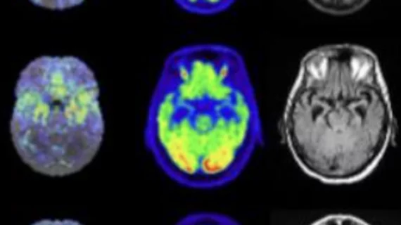
Molecular imaging (also called nuclear medicine or nuclear imaging) can image the function of cells inside the body at the molecular level. This includes the imaging modalities of positron emission computed tomography (PET) and single photon emission computed tomography (SPECT) imaging. How does PET and SPECT imaging work? Small amounts of radioactive material (radiopharmaceuticals) injected into a patient. These can use sugars or chemical traits to bond to specific cells. The radioactive material is taken up by cells that consume the sugars. The radiation emitted from inside the body is detected by photon detectors outside the body. Computers take the data to assemble images of the radiation emissions. Nuclear images may appear fuzzy or ghostly rather than the sharper resolution from MRI and CT.  But, it provides metabolic information at a cellular level, showing if there are defects in the function of the heart, areas of very high metabolic activity associated with cancer cells, or areas of inflammation, data not available from other modalities. These noninvasive imaging exams are used to diagnose cancer, heart disease, Alzheimer’s and Parkinson’s disease, bone disorders and other disorders. 

New GE HealthCare partnership expands access to prostate cancer imaging agent

The product, which is used to detect metastatic or recurrent prostate cancer, was approved by the FDA in 2021. 

Example of the four types of breast tissue density. The density of fibroglandular tissue inside the breast impacts the ability to easily see cancers. Cancers are very easy to spot in fatty breasts, but are almost impossible to find in extremely dense breasts. These examples show craniocaudal mammogram findings characterized as almost entirely fatty (far left), scattered areas of fibroglandular density (second from left), heterogeneously dense (second from right), and extremely dense (far right). RSNA

Molecular imaging ups invasive cancer detection in dense breast tissue, spots lesions missed by DBT

The modality often detects clinically important disease that is missed on standard 3D mammogram screening exams.

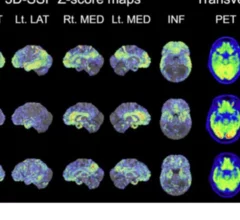
COVID-induced neuroinflammation can last 2 years or longer, according to new PET data

These findings are important to consider with regard to patients’ long-term health, as chronic inflammation has been implicated in cognitive decline. 

Thumbnail

Researchers' testing of industrial dyes could lead to the development of dementia-specific imaging agents

There are dozens of different subtypes of dementia, and it can be a challenge to determine the exact type affecting a patient. 

NIH grants to fund study of molecular imaging in stroke care

NIH is hopeful the research will enable doctors to detect signs of stroke damage earlier, opening the door for them to initiate treatments in a timelier manner.

Late-stage prostate cancer diagnoses on the rise

Experts say these new findings highlight the need for more effective screening initiatives among men who may face access barriers. 

Pharos PET scanner earns FDA clearance

Compact multifunctional PET scanner receives clearance in the US

The system was built with patients in mind, and its compact design has the potential to expand access to the modality where it may have previously been lacking. 

Fluorescent contrast agent helps diagnose basal cell carcinoma.

Fluorescent contrast agent noninvasively identifies common skin cancer

When used topically, the agent penetrates the skin and generates a fluorescent signal under the guidance of a fluorescent confocal microscope.