Molecular Imaging

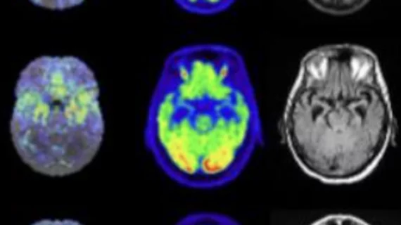
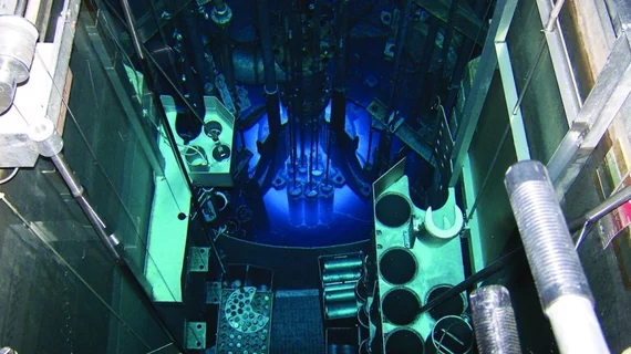
Molecular imaging (also called nuclear medicine or nuclear imaging) can image the function of cells inside the body at the molecular level. This includes the imaging modalities of positron emission computed tomography (PET) and single photon emission computed tomography (SPECT) imaging. How does PET and SPECT imaging work? Small amounts of radioactive material (radiopharmaceuticals) injected into a patient. These can use sugars or chemical traits to bond to specific cells. The radioactive material is taken up by cells that consume the sugars. The radiation emitted from inside the body is detected by photon detectors outside the body. Computers take the data to assemble images of the radiation emissions. Nuclear images may appear fuzzy or ghostly rather than the sharper resolution from MRI and CT.  But, it provides metabolic information at a cellular level, showing if there are defects in the function of the heart, areas of very high metabolic activity associated with cancer cells, or areas of inflammation, data not available from other modalities. These noninvasive imaging exams are used to diagnose cancer, heart disease, Alzheimer’s and Parkinson’s disease, bone disorders and other disorders. 

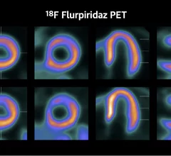
New PET radiotracer for detection of CAD approved by FDA.

New radiotracer could be a 'game changer' for diagnosing CAD

Experts are calling the agent “the most exciting development in the field of nuclear cardiology" in several decades.

Thumbnail

Novel radiotracer could improve the diagnosis of interstitial lung disease

Researchers are hopeful that uptake of the molecular SPECT imaging agent will help providers more ably monitor disease progression.

stroke brain dementia alzheimer's puzzle mental health

New tau imaging agent granted FDA's Fast Track designation

Life Molecular Imaging's new agent has demonstrated promising brain uptake, fast wash-out in non-target regions and excellent reproducibility. 

New PET technique improves imaging of pediatric neuroblastoma.

New PET technique slashes scan times, improves image quality in neuroblastoma cases

The method also reduces radiation doses and eliminates the need for sedation or general anesthesia.

FAPI PET improves staging and clinical management for a significant portion of pancreatic cancers

Fibroblast activation protein inhibitor PET imaging may provide more accurate insight into the spread of pancreatic cancer than more commonly used 18F-FDG PET/CT.

Hall of Fame Health partnership set to advance study of neurodegeneration via new PET imaging agent

The organization is teaming up with CereMark Pharma to conduct research on improving outcomes in soldiers and athletes with neurodegenerative conditions.

Thumbnail

Pharmacist pleads guilty to 'adulterating' radiopharmaceuticals

According to the DOJ, the pharmacist and those working under him “would ‘fractionate’ or ‘split’ the active ingredient of Technescan MAG3, without ensuring the pieces were equal in size, purity or strength.” 

Thumbnail

New PET method spots early signs of inflammatory bowel disease

The novel method could potentially guide decisions on when patients should start treatment.