Molecular Imaging

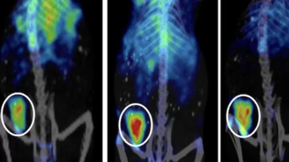
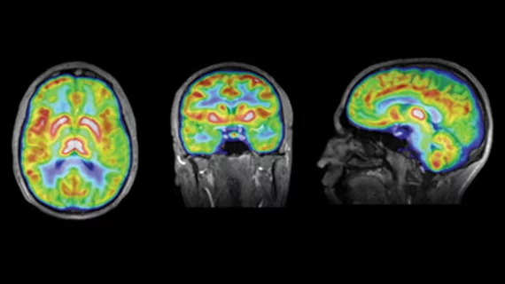
Molecular imaging (also called nuclear medicine or nuclear imaging) can image the function of cells inside the body at the molecular level. This includes the imaging modalities of positron emission computed tomography (PET) and single photon emission computed tomography (SPECT) imaging. How does PET and SPECT imaging work? Small amounts of radioactive material (radiopharmaceuticals) injected into a patient. These can use sugars or chemical traits to bond to specific cells. The radioactive material is taken up by cells that consume the sugars. The radiation emitted from inside the body is detected by photon detectors outside the body. Computers take the data to assemble images of the radiation emissions. Nuclear images may appear fuzzy or ghostly rather than the sharper resolution from MRI and CT.  But, it provides metabolic information at a cellular level, showing if there are defects in the function of the heart, areas of very high metabolic activity associated with cancer cells, or areas of inflammation, data not available from other modalities. These noninvasive imaging exams are used to diagnose cancer, heart disease, Alzheimer’s and Parkinson’s disease, bone disorders and other disorders. 

Thumbnail

World neuro-disorder survey: three out of four people would want to know

Several thousands of people were polled to see what they thought about neurological disorders. Not only did three out of four participants across 10 countries say that they would rather know if they had a disorder regardless of a cure available, but 94 percent said governments or insurers should foot the bill, according to the survey conducted by Millward Brown and announced today by survey sponsor GE Healthcare.

Anti-tau treatment blocks a form of pediatric epilepsy

A reduction in tau protein levels in the brain effectively curtails the progression of Dravet syndrome, a serious form of epilepsy in children, the Gladstone Institutes announced Aug. 13.

Thumbnail

Choline PET/MR of prostate cancer is a mismatch, but a valuable one

Quantitative values of simultaneous F-18 choline PET and diffusion-weighted imaging do not correlate in the assessment of intermediate prostate cancer—perhaps because the measurements characterize different tumor biology, according to a study published last month in PLOS ONE.

Pentagon provides $20M boon for TBI biomarkers

Abbott Laboratories based in Abbott Park, Ill., is receiving more than $19.5 million for the development of diagnostic biomarkers and assays for traumatic brain injury (TBI), the U.S. Department of Defense announced Aug. 13.

Thumbnail

A potential biomarker for PTSD

It may seem like an unlikely connection, but an aversion to kitty litter has led to a very interesting discovery about post-traumatic stress disorder (PTSD) that could one day lead to a diagnostic test and perhaps even a drug therapy,

Thumbnail

NorthStar and GE sign into partnership for U.S. isotopes

GE Healthcare and NorthStar Medical RadioIsotopes have signed a letter of intent for the production and distribution of an American supply of medical isotopes, including the largest staple isotope, molybdenum-99 (Mo-99), NorthStar announced today.

Thumbnail

Biosensors: Tracking cancer cell death on the trail of adenoviral gene therapy

A bioluminescence technique is being developed to monitor an adenovirus-mediated cancer gene therapy by following signs of apoptosis, or cell death, according to a study published online Aug. 13 in the journal Molecular Imaging.

Are anti-APOE Alzheimer’s therapies safe?

A New York Times piece reviews recent research on the safety of potential Alzheimer’s therapies that focus on genetic factors of the disease. APOE is a hallmark risk factor and perhaps the most common in those who progress to Alzheimer’s, and often earlier than others.