Molecular Imaging

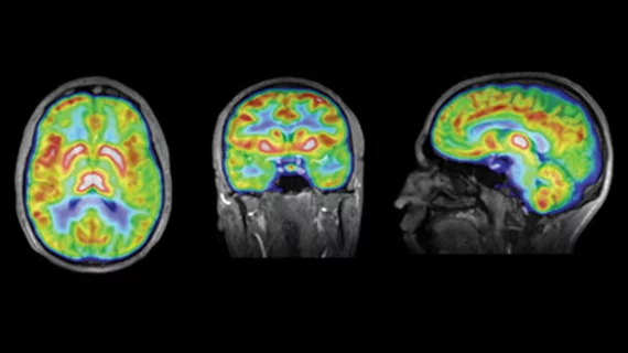
Molecular imaging (also called nuclear medicine or nuclear imaging) can image the function of cells inside the body at the molecular level. This includes the imaging modalities of positron emission computed tomography (PET) and single photon emission computed tomography (SPECT) imaging. How does PET and SPECT imaging work? Small amounts of radioactive material (radiopharmaceuticals) injected into a patient. These can use sugars or chemical traits to bond to specific cells. The radioactive material is taken up by cells that consume the sugars. The radiation emitted from inside the body is detected by photon detectors outside the body. Computers take the data to assemble images of the radiation emissions. Nuclear images may appear fuzzy or ghostly rather than the sharper resolution from MRI and CT.  But, it provides metabolic information at a cellular level, showing if there are defects in the function of the heart, areas of very high metabolic activity associated with cancer cells, or areas of inflammation, data not available from other modalities. These noninvasive imaging exams are used to diagnose cancer, heart disease, Alzheimer’s and Parkinson’s disease, bone disorders and other disorders. 

Thumbnail

Ga-68 DOTATATE PET/CT finds more NETs

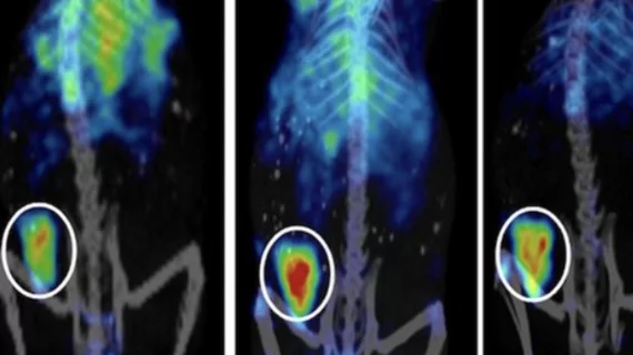
A comparison study of three different methods of neuroendocrine tumor (NET) detection that includes PET/CT, SPECT/CT and whole body MR imaging places PET/CT with gallium-68 (Ga-68) DOTATATE on a pedestal above the rest for its sensitivity in finding NETs, according to a study published Aug. 28 in the Journal of Nuclear Medicine.

Stem Cells reveal neurochemical flood in schizophrenics

Schizophrenia may be tripped off by an overabundance of chemical signals pumped out by neurons in the brain. The University of California, San Diego, announced Sept. 11 that researchers from the Skaggs School of Pharmacy and Pharmaceutical Sciences are using stem cells to see the flood of neurotransmitters first-hand.

Thumbnail

IBA and Philips to collaborate on cancer research and technology

Ion Beam Applications (IBA) announced today that the company will be teaming up with Philips Healthcare to develop new diagnostic and therapeutic research and technology, including proton therapy systems.

AAA acquires GE's FDG-PET sector in Italy

Advanced Accelerator Applications (AAA) announced today that it has acquired GE Healthcare’s FDG-PET radiopharmaceutical division in Italy.

Thumbnail

NOPR: All NaF agreements must be updated

The American College of Radiology and the Medical Imaging and Technology Alliance are prompting all participants of the bone-imaging portion of the National Oncologic PET Registry (NOPR) to sign new agreements by Sept. 22.

NanoSomix to offer Alzheimer’s blood assay for research

A blood test that predicts a patient’s likelihood of developing Alzheimer’s disease has been the target of a multi-center National Institutes of Health study and was recently publicized by the Alzheimer’s Association. Aliso Viego, Calif., based NanoSomiX announced today that it would begin providing the assay to researchers and pharmaceutical companies studying Alzheimer’s treatments this fall.

Cutting SPECT MPI scan time in half

A novel reconstruction method of cardiac SPECT myocardial perfusion imaging (MPI) could potentially reduce scan times by 50 percent and cut 75 percent of the time it takes to reconstruct images, according to research announced by the University of Eastern Finland yesterday.

Quantifying absolute MBF in dynamic SPECT

It is accepted within the cardiology community that measurements pertaining to absolute myocardial blood flow (MBF) and myocardial flow reserve (MFR) provide additional information about a patient’s heart condition than general perfusion. Researchers are still perfecting a standard procedure for obtaining these measurements, but they seem to be getting close in the case of dynamic SPECT, according to a study published Sept. 4 in the Journal of Nuclear Medicine.