Molecular Imaging

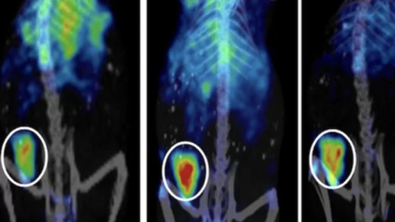
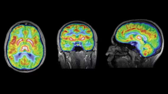
Molecular imaging (also called nuclear medicine or nuclear imaging) can image the function of cells inside the body at the molecular level. This includes the imaging modalities of positron emission computed tomography (PET) and single photon emission computed tomography (SPECT) imaging. How does PET and SPECT imaging work? Small amounts of radioactive material (radiopharmaceuticals) injected into a patient. These can use sugars or chemical traits to bond to specific cells. The radioactive material is taken up by cells that consume the sugars. The radiation emitted from inside the body is detected by photon detectors outside the body. Computers take the data to assemble images of the radiation emissions. Nuclear images may appear fuzzy or ghostly rather than the sharper resolution from MRI and CT.  But, it provides metabolic information at a cellular level, showing if there are defects in the function of the heart, areas of very high metabolic activity associated with cancer cells, or areas of inflammation, data not available from other modalities. These noninvasive imaging exams are used to diagnose cancer, heart disease, Alzheimer’s and Parkinson’s disease, bone disorders and other disorders. 

ASTRO 2014: Proton therapy's lightspeed promise

As the American Society for Radiation Oncology (ASTRO) concludes today at the Moscone Center in San Francisco, Molecular Imaging caught up with two experts in proton therapy technology and policy—David C. Beyer, MD, president-elect of ASTRO, and Sameer R. Keole, MD, vice chair of ASTRO’s government relations council and a radiation oncologist at the Mayo Clinic in Phoenix—to talk about how this cutting-edge technology is changing.

Thumbnail

Taking It to Heart: Hybrid PET for CAD

Two is better than one. At least that may prove to be the case with hybrid approaches that combine PET’s ability to assess myocardial blood flow with other modalities for diagnosing coronary artery disease.

Thumbnail

AAIC 2014: Alzheimer’s Risk Factors in Sharper Relief

A number of factors are standing out as promoters for and protectors against the development of Alzheimer’s disease, according to research presented during the 2014 Alzheimer’s Association International Conference (AAIC) that was held July 12-17 at the Bella Center in Copenhagen, Denmark.

Thumbnail

Don’t Fear the IND Labyrinth: 5 Lessons Learned at an Academic Site

The challenge was delivered with stark clarity. “We need to establish a new approach to make it easy and affordable for our faculty to quickly transition their imaging probe research from cells and what they see in animal models into human subjects.

Thumbnail

Q&A with Barry A. Siegel, MD: SNMMI Cassen Prize Winner

As the 2014 Annual Meeting for the Society of Nuclear Medicine and Molecular Imaging (SNMMI) came to a close this year in St. Louis, Molecular Imaging Insight sat down with Barry A. Siegel, MD, professor of radiology and medicine and chief of the nuclear medicine division at local Washington University School of Medicine. 

Thumbnail

Leaders in Research & Scientific Sessions

There are a number of perennial destinations for the molecular imaging set that deliver cutting-edge research and promote clinical discussion, but, for many clinicians and researchers, the Society of Nuclear Medicine and Molecular Imaging’s Annual Meeting is the one conference they attend all year.

Thumbnail

RIT takes on NHL

Advanced low-grade non-Hodgkin lymphoma (NHL) typically leads to relapse and death in patients who undergo conventional chemo and radiation therapies, but the emergence of radioimmunotherapy (RIT) in recent years has provided an encouraging alternative.

Thumbnail

MR in Hyper-drive: Introducing Hyperpolarized MRI & A New Era of Metabolic Imaging

Hyperpolarized MRI, also known as hyperpolarized MRSI (or HP-MRSI), is a new technique representing a major development in molecular MR currently making the rounds in the international molecular imaging circuit.