Molecular Imaging

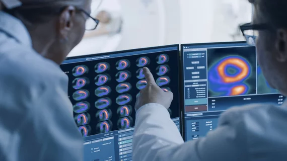
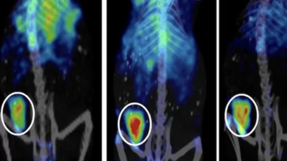
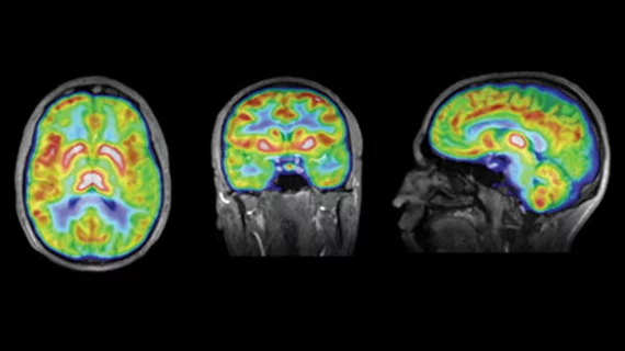
Molecular imaging (also called nuclear medicine or nuclear imaging) can image the function of cells inside the body at the molecular level. This includes the imaging modalities of positron emission computed tomography (PET) and single photon emission computed tomography (SPECT) imaging. How does PET and SPECT imaging work? Small amounts of radioactive material (radiopharmaceuticals) injected into a patient. These can use sugars or chemical traits to bond to specific cells. The radioactive material is taken up by cells that consume the sugars. The radiation emitted from inside the body is detected by photon detectors outside the body. Computers take the data to assemble images of the radiation emissions. Nuclear images may appear fuzzy or ghostly rather than the sharper resolution from MRI and CT.  But, it provides metabolic information at a cellular level, showing if there are defects in the function of the heart, areas of very high metabolic activity associated with cancer cells, or areas of inflammation, data not available from other modalities. These noninvasive imaging exams are used to diagnose cancer, heart disease, Alzheimer’s and Parkinson’s disease, bone disorders and other disorders. 

Thumbnail

Leaders in Research & Scientific Sessions

There are a number of perennial destinations for the molecular imaging set that deliver cutting-edge research and promote clinical discussion, but, for many clinicians and researchers, the Society of Nuclear Medicine and Molecular Imaging’s Annual Meeting is the one conference they attend all year.

Thumbnail

RIT takes on NHL

Advanced low-grade non-Hodgkin lymphoma (NHL) typically leads to relapse and death in patients who undergo conventional chemo and radiation therapies, but the emergence of radioimmunotherapy (RIT) in recent years has provided an encouraging alternative.

Thumbnail

MR in Hyper-drive: Introducing Hyperpolarized MRI & A New Era of Metabolic Imaging

Hyperpolarized MRI, also known as hyperpolarized MRSI (or HP-MRSI), is a new technique representing a major development in molecular MR currently making the rounds in the international molecular imaging circuit.

Thumbnail

Unlocking The Cells: Visualizing Mental Illness Via Molecular Imaging

Molecular imaging of psychiatric disorders has taken a few turns around the lab as a concept, and while some potential imaging methods, namely dopamine transport scanning, have shown some potential, the technique has not really taken off for psychiatric applications due to a mixed bag of results and a limited understanding of the pathophysiology.

Thumbnail

Rising to the Occasion: Radionuclide Therapy

First-order treatment of cancer typically involves chemo and/or radiation therapy and surgical intervention, but radionuclide therapies offer some advantages for select patients by delivering a very intense dose sequestered to affected tumor sites.

Is Molecular Imaging Due for a Fallow Period?

During the past five years, I have spent a considerable amount of time on the issue of supply of Tc-99m and its parent, Mo-99.

First-in-human study of novel integrin receptor PET tracer

The first PET imaging study of its kind to show the viability of tumor angiogenesis imaging in humans with investigational Ga-68 NODAGA-Theranost has been conducted by researchers from the Theranostics Center for Molecular Radiotherapy and Molecular Imaging in Bad Berka, Germany, and other institutions including the National Cancer Institute and Advanced Imaging Projects based in Boca Raton, Fla. Molecular Imaging has obtained an exclusive sneak-peek at the study.

Video lecture: Nanoconstructs in theranostics

Watch a presentation on nanoconstructs in cancer theranostics from the Methodist Hospital Research Institute and Tel Aviv University (TAU) posted online from the recent Summer School on Nanomedicine and Innovation.