Molecular Imaging

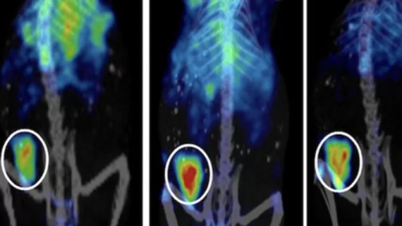
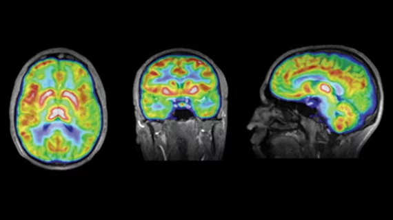
Molecular imaging (also called nuclear medicine or nuclear imaging) can image the function of cells inside the body at the molecular level. This includes the imaging modalities of positron emission computed tomography (PET) and single photon emission computed tomography (SPECT) imaging. How does PET and SPECT imaging work? Small amounts of radioactive material (radiopharmaceuticals) injected into a patient. These can use sugars or chemical traits to bond to specific cells. The radioactive material is taken up by cells that consume the sugars. The radiation emitted from inside the body is detected by photon detectors outside the body. Computers take the data to assemble images of the radiation emissions. Nuclear images may appear fuzzy or ghostly rather than the sharper resolution from MRI and CT.  But, it provides metabolic information at a cellular level, showing if there are defects in the function of the heart, areas of very high metabolic activity associated with cancer cells, or areas of inflammation, data not available from other modalities. These noninvasive imaging exams are used to diagnose cancer, heart disease, Alzheimer’s and Parkinson’s disease, bone disorders and other disorders. 

Thumbnail

Newly discovered gene linked to neurodegeneration

A gene that mediates the scale of the hippocampus in both preclinical and human brains is closely associated with the pathology of a group of neurodegenerative diseases, the University of Manchester announced yesterday.

Thumbnail

A 3D brain SPECT could lead to better cognitive function in psychiatric patients

Using 3D brain SPECT scans to guide treatment of psychiatric disorders improved health outcomes and cognitive performance, according to a study published in advance of the Winter 2014 issue of the Journal of Neuropsychiatry and Clinical Neurosciences.

FDA may ease approval for breast cancer therapeutics

A new guidance document released by the FDA on Monday provides the means to accelerate the process for approving neoadjuvant therapeutic drugs for early-stage, high-risk breast cancer.

Ohio State cuts ribbon on $13M molecular imaging facility

Ohio State University announced today that it has completed construction and development on a $13 million molecular imaging research center called Translational Research Center for Molecular Imaging Pharmaceuticals at the Columbus-based Wright Center of Innovation in Biomedical Imaging.

Thumbnail

Nobel Prize honors developers of ultra-optical microscopy

Key scientists behind the development of a prototype optical microscope have garnered the 2014 Nobel Prize in chemistry due to the technology’s ability to surpass all previous limits of optical imaging, the National Institutes of Health (NIH) announced today.

Thumbnail

FDA draws up new regulatory strategy for research, pharmaceuticals and devices

Almost 50 percent of devices and 40 percent of medications administered in the U.S. are produced outside its borders. Around 80 percent of the producers of active pharmaceutical ingredients are internationally based and the speed of global research and commerce is swiftly changing. Clearly, the regulatory framework needs to keep up. With that in mind, the FDA is setting new priorities for the coming years. An official document has been drawn up by the FDA to that effect.

Thumbnail

PET/CT changed diagnosis in 21% of breast cancer patients under 40

Patients less than 40 years of age originally diagnosed with one of the first three stages of breast cancer underwent a change in clinical staging as a result of PET/CT scanning, according to a study announced Oct. 1 by the Society of Nuclear Medicine and Molecular Imaging (SNMMI).

Thumbnail

NIR optical brain scanner wins $3.7M Marine Corps contract

A brain scanner based on differential near-infrared (NIR) energy is getting a healthy push forward with a $3.7 million, four-year contract with the U.S. Marine Corps and Navy for the development of diagnostic imaging for traumatic brain injury and hematoma, makers InfraScan announced Oct. 1.