Molecular Imaging

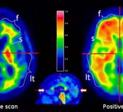
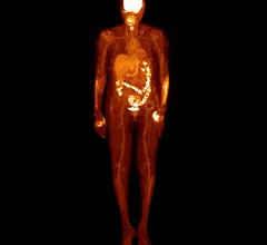
Molecular imaging (also called nuclear medicine or nuclear imaging) can image the function of cells inside the body at the molecular level. This includes the imaging modalities of positron emission computed tomography (PET) and single photon emission computed tomography (SPECT) imaging. How does PET and SPECT imaging work? Small amounts of radioactive material (radiopharmaceuticals) injected into a patient. These can use sugars or chemical traits to bond to specific cells. The radioactive material is taken up by cells that consume the sugars. The radiation emitted from inside the body is detected by photon detectors outside the body. Computers take the data to assemble images of the radiation emissions. Nuclear images may appear fuzzy or ghostly rather than the sharper resolution from MRI and CT.  But, it provides metabolic information at a cellular level, showing if there are defects in the function of the heart, areas of very high metabolic activity associated with cancer cells, or areas of inflammation, data not available from other modalities. These noninvasive imaging exams are used to diagnose cancer, heart disease, Alzheimer’s and Parkinson’s disease, bone disorders and other disorders. 

PSMA agents pinpoint prostate cancer throughout the body with SPECT, planar imaging

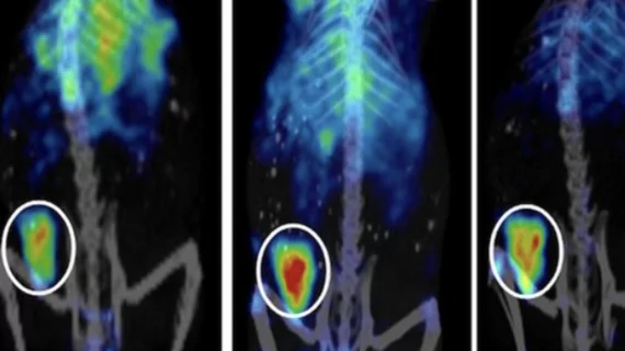
Two novel technetium-99m agents, when used with SPECT or planar imaging, have been found to effectively target prostate specific membrane antigen (PSMA) in advanced prostate cancer within bone, lymph nodes and soft tissues, according to a study published in the November issue of the Journal of Nuclear Medicine.

Thumbnail

FDG PET reigns for diagnosing infected prosthetic heart valves

Infections related to heart valve prosthesis are usually diagnosed with echocardiography, but it can miss key areas of infection. It is here that FDG PET or leukocute scintigraphy can step in, but a recent comparison study of the two nuclear medicine procedures published Nov. 13 in the Journal of Nuclear Medicine shows that FDG PET may be the best option.

Thumbnail

NorthStar snags additional $5.2M from NNSA for isotope technology

The U.S. Department of Energy’s National Nuclear Security Administration (NNSA) has agreed to provide supplemental funding for NorthStar Medical Radioisotopes’ medical isotope production venture.

Thumbnail

Canadian Light Source makes first shipment of medical isotopes from linear accelerator

The Saskatoon, Canada-based Canadian Light Source has reached a major benchmark in the company’s Medical Isotope Project by shipping the first major supply of isotopes, the company announced Friday. Health Canada has yet to approve it, but, with further testing, this technology could begin supplying medical isotopes to medical facilities in the region by 2016.

First-in-human trial of new serotonin receptor agent

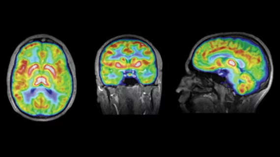
F-18 mefway is a novel serotonin-1A receptor ligand that could one day be used to image neurodegenerative disease and psychiatric disorders, according to a story published Nov. 13 in the Journal of Nuclear Medicine.

Thumbnail

Lantheus reports significant third quarter growth

Lantheus Medical Imaging, based in Billerica, Mass., announced the radiopharmaceutical company’s third quarter financials yesterday, revealing an 8 percent increase over the same quarter in 2013.

Thumbnail

New 2015 HOPPS ruling cuts into PET payments

The Centers for Medicare & Medicaid Services (CMS) issued final rules for the 2015 Hospital Outpatient Prospective Payment System (HOPPS) and the Medicare Physician Fee Schedule (MPFS). The rules were recorded in the Federal Register Nov. 10 and go into effect Jan. 1, 2015. It appears that changes to HOPPS will depress PET payments by 1.9 percent.

New blood test for Alzheimer's disease

Yet another blood test for Alzheimer's disease is being developed--this time by a group of Japanese researchers. The technique could, if approved, be used to screen patients who show no signs of neurodegeneration.