Molecular Imaging

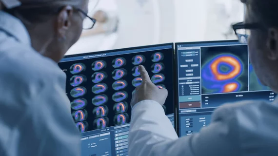
Molecular imaging (also called nuclear medicine or nuclear imaging) can image the function of cells inside the body at the molecular level. This includes the imaging modalities of positron emission computed tomography (PET) and single photon emission computed tomography (SPECT) imaging. How does PET and SPECT imaging work? Small amounts of radioactive material (radiopharmaceuticals) injected into a patient. These can use sugars or chemical traits to bond to specific cells. The radioactive material is taken up by cells that consume the sugars. The radiation emitted from inside the body is detected by photon detectors outside the body. Computers take the data to assemble images of the radiation emissions. Nuclear images may appear fuzzy or ghostly rather than the sharper resolution from MRI and CT.  But, it provides metabolic information at a cellular level, showing if there are defects in the function of the heart, areas of very high metabolic activity associated with cancer cells, or areas of inflammation, data not available from other modalities. These noninvasive imaging exams are used to diagnose cancer, heart disease, Alzheimer’s and Parkinson’s disease, bone disorders and other disorders. 

Thumbnail

Alzheimer’s: Untangling the Research

First came the discovery of beta-amyloid plaque as a precursor of cognitive decline, and then came the tangles of tau and wonderment in researchers’ minds as to their relationship.

Thumbnail

Cardiac PET/CT: Measuring calcification via low-dose CT

Low-dose CT has become a mainstay for PET attenuation correction, but why not take it a step further by quantifying coronary artery calcium without adding any extra dose, according to an Ottawa Heart Institute study published ahead of print Nov. 20 in the Journal of Nuclear Cardiology.

A Victory We Aren’t Celebrating—Yet

During my term as president of the Society of Nuclear Medicine (And Molecular Imaging, now appended), we were in the midst of the Molecular Imaging Campaign that was successful in increasing the visibility of nuclear medicine as a measure of physiologic and metabolic parameters in diagnosing our patients.

AAA financials show more than 28% spike in sales

A year-to-date sales report for the radiopharmaceutical company Advanced Accelerator Applications (AAA) indicates that the company experienced 28.9 percent growth in sales, equaling about $68.7 million in the first nine months of 2014. Net income for the first three quarters of the year was gauged at approximately $1.5 million.

Thumbnail

Capturing minute changes in DNA could aid early detection of breast cancer

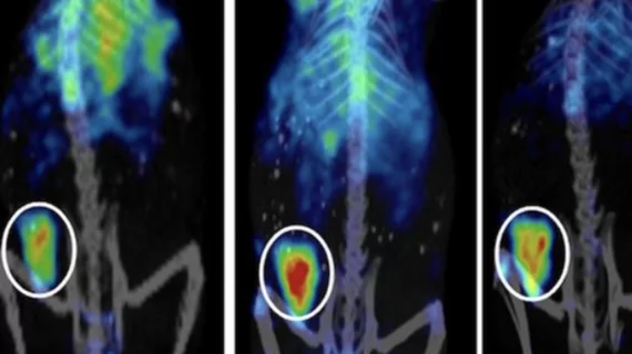
Targeting oncogene-driven activation of DNA damage could be an effective way to tap into the earliest stages of cancer development, or tumorigenesis. An investigative molecular imaging agent was able to do just that in a preclinical Oxford University study published ahead of print Nov. 13 in the Journal of Nuclear Medicine.

Thumbnail

Lymphoseek gains EU approval for sentinel node mapping in select cancers

Navidea Biopharmaceuticals announced today that technetium-99m tilmanocept (Lymphoseek) has been approved by the European Commission for the detection of sentinel lymph node involvement in primary breast cancer, melanoma and localized squamous cell carcinoma.

Thumbnail

Protea’s gross revenues up 190% in third quarter

Protea Biosciences, a biomedical company based in Morgantown, W. Va, announced yesterday that third quarter financial results amounted to $517,569, the highest revenue on record for the company. However net losses are still steep at more than $4.6 million for the third quarter.

Thumbnail

Brain PET: Periodontal disease could be a culprit in amyloid burden

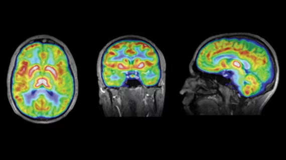
Even mild cases of dental disease could have long-term effects on amyloid plaque development and brain health, according to a neuroimaging study published online Nov. 5 in Neurobiology of Aging.