Molecular Imaging

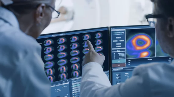
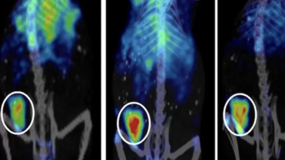
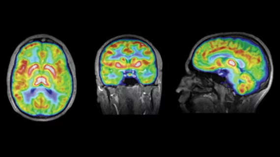
Molecular imaging (also called nuclear medicine or nuclear imaging) can image the function of cells inside the body at the molecular level. This includes the imaging modalities of positron emission computed tomography (PET) and single photon emission computed tomography (SPECT) imaging. How does PET and SPECT imaging work? Small amounts of radioactive material (radiopharmaceuticals) injected into a patient. These can use sugars or chemical traits to bond to specific cells. The radioactive material is taken up by cells that consume the sugars. The radiation emitted from inside the body is detected by photon detectors outside the body. Computers take the data to assemble images of the radiation emissions. Nuclear images may appear fuzzy or ghostly rather than the sharper resolution from MRI and CT.  But, it provides metabolic information at a cellular level, showing if there are defects in the function of the heart, areas of very high metabolic activity associated with cancer cells, or areas of inflammation, data not available from other modalities. These noninvasive imaging exams are used to diagnose cancer, heart disease, Alzheimer’s and Parkinson’s disease, bone disorders and other disorders. 

Preclinical cancer therapy leads to shrinking neuroblastoma

A cell protein known as MYCN (v-myc avian myelocytomatosis oncogene), one of a group of proteins that spur cellular proliferation, is known to be very resistant to even targeted drug treatments, but preclinical research is turning this protein on itself to fight off new neuroblastoma cancer growth, the Dana Farber Cancer Center and Boston Children’s Hospital announced today.

Thumbnail

Molecule could identify and aid treatment of aggressive breast cancers

A molecule has been found that could be leveraged to treat the most dangerous cancers of the breast, known as triple negative breast cancer, according to a Nov. 3 announcement from the City College of New York (CCNY).

Thumbnail

Grappling with the genomics of Ebola

Researchers are struggling to lock in genetic analysis related to the genetic mutability of Ebola due to delayed and incomplete data from around the world, according to a review published online today in Science.

Thumbnail

Brain DNA methylation in key genes linked to Alzheimer’s

The methylation cycles of several genetic variants—28 in fact—are associated with the hallmark neurodegeneration involved in Alzheimer’s disease (AD). Some variants were found to be associated with amyloid load and others with taupathy, according to a study published online Nov. 3 in JAMA Neurology.

FDG PET superior to SPECT for telling Alzheimer’s from DLB

Dementia imaging with FDG PET was more accurate, sensitive and specific in a comparison between Alzheimer’s disease (AD) and dementia with Lewy bodies (DLB), according to a study published online Nov. 5 in the Journal of Nuclear Medicine.

Definiens acquired by AstraZeneca and MedImmune

Global biologics company AstraZeneca and its research and development division, MedImmune, announced yesterday that the company has acquired Definiens, a biotech firm focusing on imaging and research analysis.

Thumbnail

U.K. to gain a major brain imaging center

Cardiff University in Wales has broken ground on a $70 million brain imaging center expected to be "unique within Europe." A range of brain mapping techniques will be implemented to improve researchers' understanding of diseases like multiple schlerosis, schizophrenia and dementia.

Lantheus makes deal with Shine for Mo-99 supply

Lantheus Medical announced Monday that the international radiopharmaceutical company has entered an agreement with Wisconsin-based Shine Medical Technologies for supply of molybdenum-99 (Mo-99).