Molecular Imaging

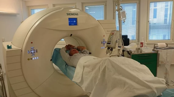
Molecular imaging (also called nuclear medicine or nuclear imaging) can image the function of cells inside the body at the molecular level. This includes the imaging modalities of positron emission computed tomography (PET) and single photon emission computed tomography (SPECT) imaging. How does PET and SPECT imaging work? Small amounts of radioactive material (radiopharmaceuticals) injected into a patient. These can use sugars or chemical traits to bond to specific cells. The radioactive material is taken up by cells that consume the sugars. The radiation emitted from inside the body is detected by photon detectors outside the body. Computers take the data to assemble images of the radiation emissions. Nuclear images may appear fuzzy or ghostly rather than the sharper resolution from MRI and CT.  But, it provides metabolic information at a cellular level, showing if there are defects in the function of the heart, areas of very high metabolic activity associated with cancer cells, or areas of inflammation, data not available from other modalities. These noninvasive imaging exams are used to diagnose cancer, heart disease, Alzheimer’s and Parkinson’s disease, bone disorders and other disorders. 

Radiocholine PET/CT has a niche in prostate imaging

Selection of patients with advanced prostate cancer who are eligible for salvage radiation therapy could be improved with the use of radiocholine PET/CT, according to a review of recent research published today in the Journal of Nuclear Medicine.

Not just three scans: Understanding FDG PET coverage

The Centers for Medicare & Medicaid Services provided a reiteration of the agency’s national coverage decision regarding oncologic FDG PET this summer and clarified specifically the –KX modifier that needs to be used when clinicians code for the fourth scan and beyond.

Thumbnail

Neuro study of worms is a 'brain hack'

Nematode worms are providing a very, very small window on the neural connections that we humans share. A feature in New Scientist follows the circuitous path of our knowledge about consciousness and the neural networks of the brain.

Plumbing the depths of PTSD

A feature in The New Yorker explores how Daniela Schiller, PhD, from the Mount Sinai School of Medicine’s Brain Imaging Core in New York City, has approached a kind of memory erasure for people who have suffered greatly from post-traumatic stress disorder (PTSD) with the aid of cutting-edge neuroimaging.

Consortium to develop MR-guided radiotherapy

The Christie NHS Foundation Trust in Manchester, U.K., is the newest institution to join an assembly of organizations with the objective of researching an integrated and real-time MR-guided radiotherapy system for more accurate treatment delivery, according to today’s announcement from Elekta and Philips Healthcare.

Thumbnail

Zinc imaging could be used to track a spate of diseases

A preliminary imaging study with Zn-63 zinc citrate confirmed a protocol for preparation and showed encouraging preclinical biodistribution. This technique holds promise for a wide variety of diseases involving zinc disorder, including a range of cancers and metabolic disorders, according to a study published July 21 in the Journal of Nuclear Medicine.

FDA: Shortfall of Ceretec technetium kit

The FDA announced July 14 that a shortage of Technetium Tc99m Exametazime Injection, also known as the Ceretec Kit (GE Healthcare, Medi-Physics), is in effect and expected to continue until the first week of August.

Thumbnail

TDP-43: Yet another protein implicated in Alzheimer's

Alzheimer's disease is characterized by the hallmark pathology of beta-amyloid plaque and tau protein tangles, but there appears to be a new protein on the block in the development of the neurodegenerative disease. It is called TAR DNA binding protein of 43kDa (TDP-43), according to research presented during the 2014 international conference of the Alzheimer's Association (AAIC).