Molecular Imaging

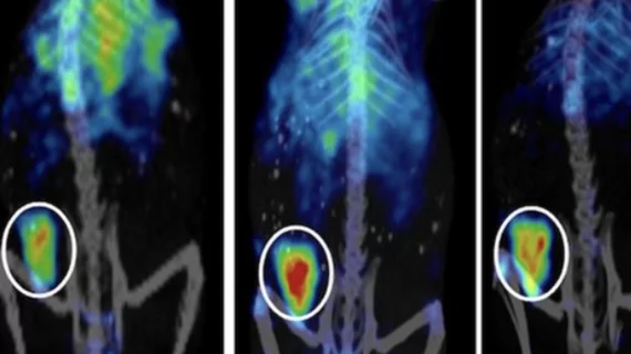
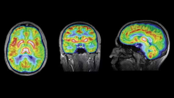
Molecular imaging (also called nuclear medicine or nuclear imaging) can image the function of cells inside the body at the molecular level. This includes the imaging modalities of positron emission computed tomography (PET) and single photon emission computed tomography (SPECT) imaging. How does PET and SPECT imaging work? Small amounts of radioactive material (radiopharmaceuticals) injected into a patient. These can use sugars or chemical traits to bond to specific cells. The radioactive material is taken up by cells that consume the sugars. The radiation emitted from inside the body is detected by photon detectors outside the body. Computers take the data to assemble images of the radiation emissions. Nuclear images may appear fuzzy or ghostly rather than the sharper resolution from MRI and CT.  But, it provides metabolic information at a cellular level, showing if there are defects in the function of the heart, areas of very high metabolic activity associated with cancer cells, or areas of inflammation, data not available from other modalities. These noninvasive imaging exams are used to diagnose cancer, heart disease, Alzheimer’s and Parkinson’s disease, bone disorders and other disorders. 

Thumbnail

Amino-acid PET imaging of gliomas in the spotlight

Amino-acid biomarkers have undergone an evolution in the imaging of gliomas. Several agents have shown to be particularly useful, most notably C-11 MET, F-18 FDOPA, FLT and FET, according to a review of amino-acid imaging published July 8 in the Journal of Nuclear Medicine.

FET-PET may monitor treatment response in glioblastoma

Preliminary research shows that O-(2-[18F]-fluoroethyl)-L-tyrosine (FET) could be used as a biomarker for response to chemoradiotherapy in glioblastoma patients, the University of West Australia School of Medicine announced last week.

Novel optical imaging system tracks toxicity via DNA accumulation

A preclinical imaging system that takes conventional microscopy and transforms it into a molecular imaging platform uses fluorescence and luminescence as well as radioisotope biomarkers and high resolution x-ray. The technique is called intravital microscopy, according to a study published online July 12 in Hepatology.

AAIC 2014 convenes in Copenhagen

The Alzheimer's Association International Conference is schedule to be held July 12-17 at the Bella Center in Copenhagen, Denmark. The meeting will feature many scientific sessions, plenary sessions and symposia dedicated to the most up-to-date Alzheimer's disease research.

Thumbnail

Data uncover genomic signatures of most common lung cancer

Specific genetic mutations in an ear-marked pathway now have been implicated in the development of lung adenocarcinoma, the most prevalent subtype of lung cancer, according to a study published online July 9 in Nature. The discovery of these mutations could hone drug targets for new therapies down the road.

Patients at Henry Ford Wyandotte now receive half-dose nuclear medicine imaging

UltraSpect, provider of the only multi-vendor and most cost-effective imaging solution for meeting the American Society of Nuclear Cardiology (ASNC) low dose guidelines, announces today that Henry Ford Wyandotte Hospital in Michigan has upgraded its nuclear medicine (NM) image reconstruction software to UltraSpect Xpress3.Cardiac. As a result, all patients receiving NM exams now have exams in half the time, with half the dose, regardless of the model or age of camera being used.

Thumbnail

AAA financials reveal 30% uptick in sales

An annual financial report from Advanced Accelerator Applications (AAA) in Saint-Genis-Pouilly, France, announced yesterday that the company’s sales were up just over 30 percent to almost $80 million.

Protea and InSphero collaborate on 3D preclinical imaging

Protea Biosciences based in West Virginia and Swiss biotech firm InSphero announced this week that the companies are combining their 3D technologies into one preclinical imaging platform.