Molecular Imaging

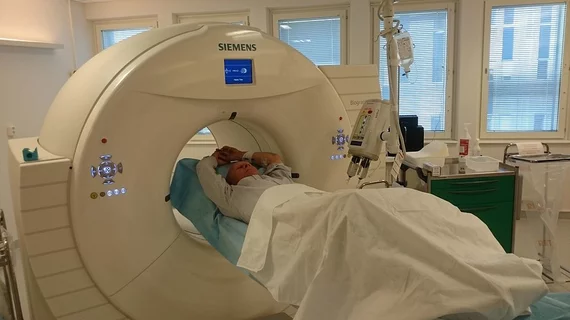
Molecular imaging (also called nuclear medicine or nuclear imaging) can image the function of cells inside the body at the molecular level. This includes the imaging modalities of positron emission computed tomography (PET) and single photon emission computed tomography (SPECT) imaging. How does PET and SPECT imaging work? Small amounts of radioactive material (radiopharmaceuticals) injected into a patient. These can use sugars or chemical traits to bond to specific cells. The radioactive material is taken up by cells that consume the sugars. The radiation emitted from inside the body is detected by photon detectors outside the body. Computers take the data to assemble images of the radiation emissions. Nuclear images may appear fuzzy or ghostly rather than the sharper resolution from MRI and CT.  But, it provides metabolic information at a cellular level, showing if there are defects in the function of the heart, areas of very high metabolic activity associated with cancer cells, or areas of inflammation, data not available from other modalities. These noninvasive imaging exams are used to diagnose cancer, heart disease, Alzheimer’s and Parkinson’s disease, bone disorders and other disorders. 

NorthStar Medical teams with Triad for domestic supply of moly

NorthStar Medical Radioisotopes, based in Madison, Wis., announced a non-exclusive letter of intent yesterday with major radiopharmacy chain Triad Isotopes in an effort to provide a U.S. source of molybdenum-99 (Mo-99), which decays to produce technetium-99m (Tc-99m), used in the lion's share of nuclear medicine procedures.

FDA approves Lymphoseek to help determine the extent of head and neck cancer in the body

The U.S. Food and Drug Administration today approved a new use for Lymphoseek (technetium 99m tilmanocept) Injection, a radioactive diagnostic imaging agent used to help doctors determine the extent a type of cancer called squamous cell carcinoma has spread in the body’s head and neck region.

GE Healthcare Launches Interactive Patient Profiler for AdreView (Iobenguane I 123 Injection)

The Interactive Patient Profiler for AdreView is a simulation tool designed to help educate physicians on the use of AdreView (Iobenguane I 123 Injection) to assess mortality risk in patients with heart failure. The tool demonstrates the types of patients where the use of AdreView can provide further information to assist physicians in identifying patients who may have a lower one- and two-year mortality risk, among groups of patients with similar profiles based on other clinical information.

SNMMI 2014: Interview with Barry A. Siegel, MD, winner of Cassen Prize

As the 2014 Annual Meeting for the Society of Nuclear Medicine and Molecular Imaging (SNMMI) came to a close this year, Molecular Imaging sat down with Barry A. Siegel, MD, professor of radiology and medicine and chief of the nuclear medicine division at Washington University School of Medicine here in St. Louis. Siegel was honored this year with the most prestigious award presented during the meeting: the Benedict Cassen Prize.

MAA price spike: Jubilant DraxImage address dodges antitrust concern

General discontent followed a one-time, exponential price hike from Jubilant DraxImage starting April 1 for the company’s exclusive MAA kit for the preparation of technetium-99m albumin, a lung perfusion agent. An industry forum on challenges in the radiopharmaceutical realm presented during the recent Society of Nuclear Medicine and Molecular Imaging (SNMMI) 2014 Annual Meeting in St. Louis featured a panel of speakers, including vice president of sales, marketing and business development for JDI, Kevin Brooks, who made a case for the company.

Research on Piramal Imaging's Neuraceq (florbetaben F18 injection) takes center stage at SNMMI

Piramal Imaging released details from research presented at the Annual Meeting of the Society of Nuclear Medicine and Molecular Imaging (SNMMI), currently underway at St. Louis Convention Center in St. Louis, Mo.

Thumbnail

Amyloid versus tau?

It is widely publicized that amyloid in the brain may not be the best agent for gauging progression and severity of Alzheimer’s disease. Tau imaging seems to hold greater promise in that arena. What does this mean for amyloid imaging?

Thumbnail

Novel HER3 biomarker for advanced breast cancer could lead to new therapies

Human epidermal growth factor receptor 2 (HER2) biomarkers have been the standard for imaging agents that target hormonally active breast cancers that tend to be very aggressive. Now researchers have developed an alternative and potentially more comprehensive biomarker of disease, HER3, according to a study presented during this week’s Society of Nuclear Medicine and Molecular Imaging’s 2014 Annual Meeting, which was held June 7-11 in St. Louis.