Molecular Imaging

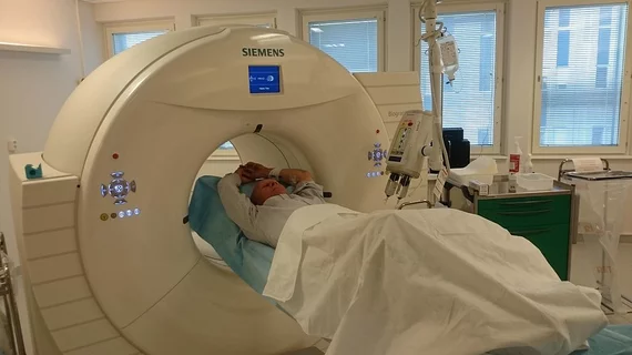
Molecular imaging (also called nuclear medicine or nuclear imaging) can image the function of cells inside the body at the molecular level. This includes the imaging modalities of positron emission computed tomography (PET) and single photon emission computed tomography (SPECT) imaging. How does PET and SPECT imaging work? Small amounts of radioactive material (radiopharmaceuticals) injected into a patient. These can use sugars or chemical traits to bond to specific cells. The radioactive material is taken up by cells that consume the sugars. The radiation emitted from inside the body is detected by photon detectors outside the body. Computers take the data to assemble images of the radiation emissions. Nuclear images may appear fuzzy or ghostly rather than the sharper resolution from MRI and CT.  But, it provides metabolic information at a cellular level, showing if there are defects in the function of the heart, areas of very high metabolic activity associated with cancer cells, or areas of inflammation, data not available from other modalities. These noninvasive imaging exams are used to diagnose cancer, heart disease, Alzheimer’s and Parkinson’s disease, bone disorders and other disorders. 

SNMMI 2014: Circulating cancer cells count down to metastasis

High-powered radioluminescence microscopy can home in on extremely scarce malignant cells among billions of normal cells in a blood sample. Leveraging the molecular imaging of circulating tumor cells (CTCs) could mean early detection of metastasis and the potential to improve survival for patients with several varieties of cancer, according to a study presented today during the Society of Nuclear Medicine and Molecular Imaging (SNMMI) 2014 Annual Meeting press conference.

Thumbnail

SNMMI 2014: Welcome montage

ST. LOUIS--As the weekend opening of the 61st Society of Nuclear Medicine and Molecular Imaging (SNMMI) Annual Meeting sunsets, three more days full of meetings, presentations and exhibitions approach. See an SNMMI-hosted audiovisual montage of highlights and attendee interviews from the first two days of the conference.

Thumbnail

ImaginAb and UCLA strike a licensing agreement for immuno-PET agents

Los Angeles-based biomedical firm ImaginAb has announced June 3 that the company is working with researchers at the University of California and has settled on a technology licensing agreement for the developmentof investigational immune cell PET agents.

ImaginAb Presents Initial Phase I/IIa Results for Prostate Imaging Agent at Society of Nuclear Medicine and Molecular Imaging (SNMMI) Annual Meeting

ImaginAb, Inc., in collaboration with Memorial Sloan-Kettering Cancer Center (MKSCC), will be presenting initial clinical data with an anti-PSMA recombinant antibody fragment (a "minibody") derived from the huJ591 monoclonal antibody, in patients with metastatic prostate cancer. The presentation of this initial data coincides with the Company's preparation to expand the clinical development of this promising imaging agent in the US and internationally.

Thumbnail

Parkinsonism: Transplanted stem cells survive and thrive

Remediation of Parkinson’s via stem cell transplantation has been verified and dopamine transporter (DAT) expression was found to be normal not just in short-term studies, but for 4-14 years after transplantation of dopamine neurons, according to a study published June 5 in Cell Reports.

Thumbnail

Genetic analysis: Essential for cancer care

The new cancer center paradigm provides genetic tests not just for experimental research, but as a clinical standard. In this glimpse by Bloomberg, Memorial Sloan Kettering Cancer Center shows off a new laboratory opened just weeks ago where patients' biopsies will be tested for 341 mutations associated with disease.

ASNC to hold nuclear cardiology board exam preparation course July 2014

Bethesda, MD (June 3, 2014) The American Society of Nuclear Cardiology (ASNC) will be offering a Nuclear Cardiology Board Exam Preparation Course from July 11-13 at the Intercontinental Hotel Chicago O'hare in Rosemont, Ill.

Thumbnail

Unlocking the great cell Mediator

Using high-powered electron microscopy, scientists at the Scripps Research Institute in La Jolla, Calif., have uncovered one of the most important cellular mechanisms of gene expression, the Mediator, according to a study published today in the journal Cell. The researchers’ new map of cellular machinery is key to a deeper understanding of cellular processes, including the production of proteins.