Molecular Imaging

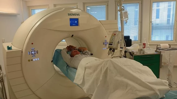
Molecular imaging (also called nuclear medicine or nuclear imaging) can image the function of cells inside the body at the molecular level. This includes the imaging modalities of positron emission computed tomography (PET) and single photon emission computed tomography (SPECT) imaging. How does PET and SPECT imaging work? Small amounts of radioactive material (radiopharmaceuticals) injected into a patient. These can use sugars or chemical traits to bond to specific cells. The radioactive material is taken up by cells that consume the sugars. The radiation emitted from inside the body is detected by photon detectors outside the body. Computers take the data to assemble images of the radiation emissions. Nuclear images may appear fuzzy or ghostly rather than the sharper resolution from MRI and CT.  But, it provides metabolic information at a cellular level, showing if there are defects in the function of the heart, areas of very high metabolic activity associated with cancer cells, or areas of inflammation, data not available from other modalities. These noninvasive imaging exams are used to diagnose cancer, heart disease, Alzheimer’s and Parkinson’s disease, bone disorders and other disorders. 

PSMA-PET/CT: New radiotracer pictures more prostate tumors

An emerging radiotracer homes in on prostate-specific anatomy and finds elusive lesions, according to a study presenting this year at the Society of Nuclear Medicine and Molecular Imaging’s 2014 Annual Meeting.

Depressed elderly may progress more rapidly to Alzheimer’s

The combination of late-life depression and beta-amyloid deposition may lead to a quickening of neurodegeneration and progressive Alzheimer’s disease, according to a study presented during the Society of Nuclear Medicine and Molecular Imaging’s 2014 Annual Meeting, which concludes today.

Thumbnail

Highlights from the 2014 SNMMI Annual Meeting

Wednesday is the last day of the Society of Nuclear Medicine and Molecular Imaging's 2014 Annual Meeting in St. Louis, but the news and images keep coming.

NorthStar Medical Technologies Signs Letter of Intent with Triad Isotopes To Bring New Domestic Source of Non-HEU Materials To Market

NorthStar Medical Technologies, LLC today announced the signing of a non-exclusive Letter of Intent with Triad Isotopes, Inc.  The agreement solidifies Triad’s position as an early adopter of domestically-sourced Non-HEU Molybdenum99 through NorthStar’s unique intelligent isotope separation systems, once that technology is approved by the FDA and commercially available.

Philips Drives Digital Innovation with Vereos at Society of Nuclear Medicine and Molecular Imaging Annual Meeting

Royal Philips today introduced Vereos PET/CT, the first digital PET/CT scanner, at the 2014 annual meeting of the Society of Nuclear Medicine and Molecular Imaging  (SNMMI), taking place this week in St. Louis, Mo.  In addition to Vereos, Philips will showcase a selection of innovative, molecular imaging solutions designed to deliver outstanding image quality, critical clinical information and greater connectivity.

Thumbnail

SNMMI 2014: Sentinel lymph node SPECT/CT spots more cancer

Society of Nuclear Medicine and Molecular Imaging (SNMMI)  leadership and a panel of experts announced today the most striking research being presented at the 2014 SNMMI Annual Meeting at the America’s Center convention center from June 7-11. This included the abstract of the year outlining how sentinel lymph node SPECT/CT imaging bests lymphoscintigraphy.

SNMMI 2014: Stem-cell treatment could halt damage from a heart attack

Acute myocardial infarction causes thousands of fatalities each year. What if there was a way to use stem-cell regeneration to prevent the devastating after-effects of a heart attack? According to researchers presenting a Japanese study at the Society of Nuclear Medicine and Molecular Imaging’s 2014 Annual Meeting, this therapy exists and could potentially prevent dangerous cardiac tissue damage.

Thumbnail

SNMMI 2014: Tau PET study takes Image of the Year

Novel PET imaging of the hallmark neurofibrillary tangles in Alzheimer’s disease using F-18 THK5117 has nabbed the Society of Nuclear Medicine and Molecular Imaging’s 2014 Image of the Year award. The study involved a head-to-head comparison of tau and amyloid PET imaging to verify whether tau PET can image the progression of Alzheimer’s pathology.