Molecular Imaging

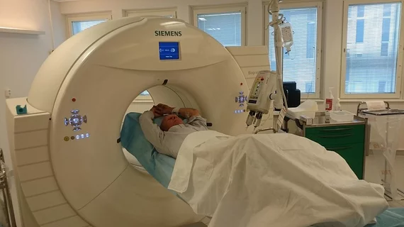
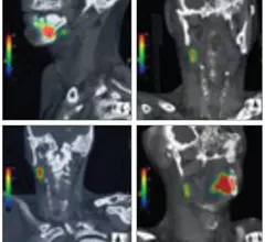
Molecular imaging (also called nuclear medicine or nuclear imaging) can image the function of cells inside the body at the molecular level. This includes the imaging modalities of positron emission computed tomography (PET) and single photon emission computed tomography (SPECT) imaging. How does PET and SPECT imaging work? Small amounts of radioactive material (radiopharmaceuticals) injected into a patient. These can use sugars or chemical traits to bond to specific cells. The radioactive material is taken up by cells that consume the sugars. The radiation emitted from inside the body is detected by photon detectors outside the body. Computers take the data to assemble images of the radiation emissions. Nuclear images may appear fuzzy or ghostly rather than the sharper resolution from MRI and CT.  But, it provides metabolic information at a cellular level, showing if there are defects in the function of the heart, areas of very high metabolic activity associated with cancer cells, or areas of inflammation, data not available from other modalities. These noninvasive imaging exams are used to diagnose cancer, heart disease, Alzheimer’s and Parkinson’s disease, bone disorders and other disorders. 

Thumbnail

Making the Mark: Incoming SNMMI President Offers a Blueprint for the Year

A knowledgeable voice among nuclear medicine leaders, the president-elect of the Society of Nuclear Medicine and Molecular Imaging (SNMMI) returns to the pages of Molecular Imaging Insight with some commentary on recent changes in the industry, new items on the molecular imaging agenda and worthy initiatives in the wings.

Thumbnail

Tau imaging: A review of the triumphs and challenges

Tau has been identified as one of the keys to unlocking neurodegenerative disease, including Alzheimer’s. However, efforts to develop an ideal tau PET agent continue to be an uphill battle, according to a review published May 15 in the Journal of Nuclear Medicine.

Pancreatic PET stress test could eliminate cumbersome testing

A minimally invasive PET scan could tell clinicians about a patient’s exocrine function without all of the usual pains of conventional testing, according to a study published May 19 in the Journal of Nuclear Medicine.

Thumbnail

Biobank launches brain study

In this video, BBC reporter Fergus Walsh is the first to undergo an MRI scan as part of the largest scan study to date and describes how the study is evolving.

IBA Molecular awarded 3-year group purchasing agreement with Premier, Inc.

IBA Molecular North America, Inc. (IBA Molecular) announced the signing of a 3-year group purchasing agreement with Premier, Inc. (Premier), a leading health care improvement company, to offer PET radiopharmaceuticals, including 18F-Fluorodeoxyglucose (FDG), a diagnostic radiotracer used in oncology, neurology and cardiology, to Premier member imaging centers across the country.

Thumbnail

World’s largest brain study focuses on dementia

An estimated 100,000 participants are expected to get involved in a major effort to understand the pathology of dementia. Medical research institute UK Biobank is planning to image 9,000 cases of Alzheimer’s disease by 2022.

MIT develops lightning fast 3D optical neuroimaging system

Based on principles of light-field microscopy, an international team of researchers has created a novel molecular imaging technique that allows three-dimensional scenes of an entire brain’s neural networks in milliseconds flat, officials from the Massachusetts Institute of Technology (MIT) announced May 19.

Stony Brook receives $3.5 million gift for new molecular imaging lab

A gift of $3.5 million has been given in a single donation to establish a molecular imaging facility at the State University of New York (SUNY), Stony Brook. Provided by Lalit and Kavita Bahl, the gifted facility will include a cyclotron and be named the Bahl Molecular Imaging Laboratory.