Molecular Imaging

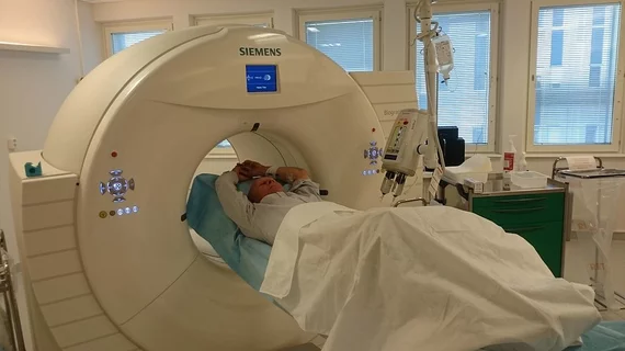
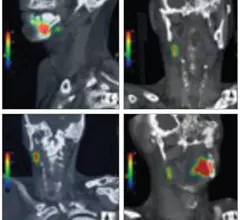
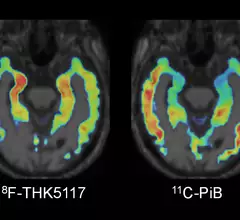
Molecular imaging (also called nuclear medicine or nuclear imaging) can image the function of cells inside the body at the molecular level. This includes the imaging modalities of positron emission computed tomography (PET) and single photon emission computed tomography (SPECT) imaging. How does PET and SPECT imaging work? Small amounts of radioactive material (radiopharmaceuticals) injected into a patient. These can use sugars or chemical traits to bond to specific cells. The radioactive material is taken up by cells that consume the sugars. The radiation emitted from inside the body is detected by photon detectors outside the body. Computers take the data to assemble images of the radiation emissions. Nuclear images may appear fuzzy or ghostly rather than the sharper resolution from MRI and CT.  But, it provides metabolic information at a cellular level, showing if there are defects in the function of the heart, areas of very high metabolic activity associated with cancer cells, or areas of inflammation, data not available from other modalities. These noninvasive imaging exams are used to diagnose cancer, heart disease, Alzheimer’s and Parkinson’s disease, bone disorders and other disorders. 

Thumbnail

Q&A with a PET Agent Expert

An exclusive interview with Michael Graham, MD, PhD, covering the latest developments in PET radiopharmaceuticals, from drugs just approved and blazing trail in clinical use, to compounds in the far reaches of research.

Thumbnail

Isotope Crisis: New Technologies Primed for Post-Chalk River Production

Continued shutdowns at research reactors throughout the world have created shortfalls in essential medical isotope supply for several years. 

Thumbnail

Strengths, Struggles & Strategies Managing Image Data

The drive for heterogeneity in molecular imaging calls for management of complex imaging data in massive volumes, a task that is challenging information technology experts and medical community members alike. 

Thumbnail

Image of Youth: Progress in Molecular Pediatric Medicine

Molecular imaging has become increasingly important in the non-invasive diagnosis and monitoring of pediatric disease, whether known or suspected. Innovations in molecular oncology, neuroimaging and hybrid imaging are shaping how clinicians approach a range of diseases in children and young adults.

Thumbnail

Advances in genetics and medical imaging meet Richard III

Shakespeare called King Richard III a hunchback, but new genetic and reconstructive CT data show this story had more of a twist. Until recently, the exact nature and whereabouts of Richard's physical deformity was a mystery. While questions still daunt researchers, a compelling picture of the monarch's spine has emerged.

PET radiopharmaceutical use jumps almost 10% in select institutions

The use of PET radiopharmaceuticals increased from 17 percent to 26 percent in diagnostic nuclear medicine procedures, according to a report evaluating adverse effects published May 29 in the Journal of Nuclear Medicine.

Thumbnail

Perfusion scintigraphy safer for pregnant women with pulmonary embolism

In a comparison safety study between scintigraphy and 256-slice angiography for pregnant women suspected of having a pulmonary embolism, scintigraphy was found to be the safest choice for moms and their unborn babies, according to a study published May 29 in the Journal of Nuclear Medicine.

Lilly invests $80M in UK biomedical and teams up with Qiagen

The biopharmaceutical company Eli Lilly based in Indianapolis, announced yesterday that it is collaborating in the development of Qiagen’s modular assay panels that allow simultaneous study of multiple biomarkers under investigation by Lilly. This comes on the heels of news that Lilly has made a sizable investment in an $80 million Epidarex Capital fund for early-stage life science and health technology companies in the United Kingdom.