Molecular Imaging

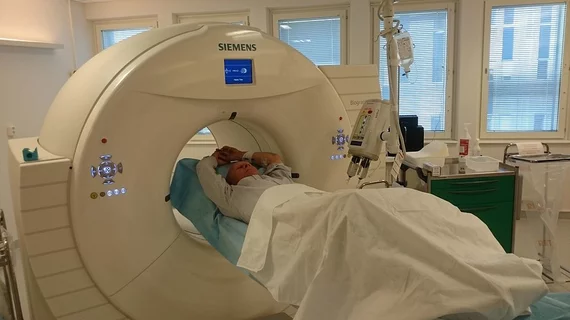
Molecular imaging (also called nuclear medicine or nuclear imaging) can image the function of cells inside the body at the molecular level. This includes the imaging modalities of positron emission computed tomography (PET) and single photon emission computed tomography (SPECT) imaging. How does PET and SPECT imaging work? Small amounts of radioactive material (radiopharmaceuticals) injected into a patient. These can use sugars or chemical traits to bond to specific cells. The radioactive material is taken up by cells that consume the sugars. The radiation emitted from inside the body is detected by photon detectors outside the body. Computers take the data to assemble images of the radiation emissions. Nuclear images may appear fuzzy or ghostly rather than the sharper resolution from MRI and CT.  But, it provides metabolic information at a cellular level, showing if there are defects in the function of the heart, areas of very high metabolic activity associated with cancer cells, or areas of inflammation, data not available from other modalities. These noninvasive imaging exams are used to diagnose cancer, heart disease, Alzheimer’s and Parkinson’s disease, bone disorders and other disorders. 

Thumbnail

World’s largest brain study focuses on dementia

An estimated 100,000 participants are expected to get involved in a major effort to understand the pathology of dementia. Medical research institute UK Biobank is planning to image 9,000 cases of Alzheimer’s disease by 2022.

MIT develops lightning fast 3D optical neuroimaging system

Based on principles of light-field microscopy, an international team of researchers has created a novel molecular imaging technique that allows three-dimensional scenes of an entire brain’s neural networks in milliseconds flat, officials from the Massachusetts Institute of Technology (MIT) announced May 19.

Stony Brook receives $3.5 million gift for new molecular imaging lab

A gift of $3.5 million has been given in a single donation to establish a molecular imaging facility at the State University of New York (SUNY), Stony Brook. Provided by Lalit and Kavita Bahl, the gifted facility will include a cyclotron and be named the Bahl Molecular Imaging Laboratory.

Thumbnail

SPECT/CT superior for spotting gastrointestinal bleeding

When compared to planar technetium-99m imaging, SPECT/CT was more adept at finding the source of acute gastrointestinal bleeds, according to a study presented May 8 during the 2014 annual meeting of the American Roentgen Ray Society held in San Diego.

International Isotopes reports 17% first quarter gain

Idaho Falls, Idaho, producer of nuclear medicine products and services International Isotopes announced May 15 the financial results for the first quarter 2014. Revenue was reported as $1,948,856, up 17 percent from $1,672,789 for the same timeframe last year.

Neuro expert Ira Shoulson joins Prana’s board of directors

Australia is a hotbed of Alzheimer’s and other neuroimaging and therapeutics research. Melbourne-based Prana Biotechnology is bringing in a new director, Ira Shoulson, MD, to serve on the company’s board.

Thumbnail

Glass Brain shows 3D detail of brain connectivity

Watch a fly-through of Glass Brain, a 3D imagining of the human brain by researchers at the University of California San Francisco. This video shows the 3D brain model made possible by electroencephalography data and white matter tract reconstruction from MRI diffusion tensor imaging. UCSF is using the technology to research possible treatments for neurodegenerative disease.

Quantitative imaging and genomics merge with CorTechs Labs and HLI database

CorTechs Labs, a quantitative imaging technology company in San Diego, is teaming up with the genomics and therapeutics group Human Longevity Inc. (HLI) to align their data into a comprehensive phenotyping database.