Molecular Imaging

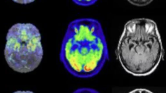
Molecular imaging (also called nuclear medicine or nuclear imaging) can image the function of cells inside the body at the molecular level. This includes the imaging modalities of positron emission computed tomography (PET) and single photon emission computed tomography (SPECT) imaging. How does PET and SPECT imaging work? Small amounts of radioactive material (radiopharmaceuticals) injected into a patient. These can use sugars or chemical traits to bond to specific cells. The radioactive material is taken up by cells that consume the sugars. The radiation emitted from inside the body is detected by photon detectors outside the body. Computers take the data to assemble images of the radiation emissions. Nuclear images may appear fuzzy or ghostly rather than the sharper resolution from MRI and CT.  But, it provides metabolic information at a cellular level, showing if there are defects in the function of the heart, areas of very high metabolic activity associated with cancer cells, or areas of inflammation, data not available from other modalities. These noninvasive imaging exams are used to diagnose cancer, heart disease, Alzheimer’s and Parkinson’s disease, bone disorders and other disorders. 

Genomic research could spell new drugs for schizophrenia and other disorders

A partnership between the Broad Institute’s Stanley Center for Psychiatric Research in Cambridge, Mass., and Tetra Discovery Partners based in Grand Rapids, Mich., that pores over the human genome for specific variants in schizophrenia may make way for new pharmaceutical therapies for psychiatric disease, Tetra announced April 9.

Thumbnail

A-synuclein SPECT agent scopes out Parkinson’s

A prospective Parkinson’s imaging agent taps into alpha-synuclein deposition in the brain, developers ICB International announced April 11.

Parkinson's gene promotes deadly protein over-production

Runaway production of proteins tripped off by a genetic mutation may be the culprit in the neurological damage associated with Parkinson’s disease, the National Institutes of Health announced yesterday.

Light-activated nanoparticle cancer drug delivery dazzles

A novel theranostics technique uses dual infrared photon laser beams to release chemotherapy within tumor cells for next-generation imaging biomarker and targeted therapy, according to an announcement from the Jonsson Comprehensive Cancer Center at the University of California, Los Angeles.

Epigenetic circuits reveal glioblastoma stem cells, potential therapy

A small group of proteins called transcription factors that regulate genetic expression tip off the mother cells of very aggressive and therapy-resistant brain cancer, according to Massachusetts General Hospital research announced by the institution yesterday.

European Commission commits $1.8M grant for intraoperative cancer imaging

An international consortium has been awarded a $1.8 million grant from the European Commission to assess Cerenkov luminescence imaging for intraoperative imaging of gastric cancer patients.

House bill champions international effort for dementia care

A bipartisan bill brought forward in the U.S. House of Representatives April 4 would coordinate an international initiative for the prevention and treatment of Alzheimer’s disease and other dementias.

Thumbnail

FDA sees through GE Q.Clear PET/CT image reconstruction

GE Healthcare announced yesterday that the U.S. Food and Drug Administration (FDA) has provided 510(k) clearance for the company’s PET/CT iterative image reconstruction technology that works to improve image quality and quantitative data.