Molecular Imaging

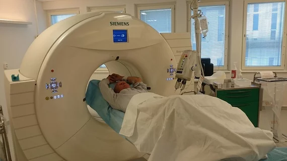
Molecular imaging (also called nuclear medicine or nuclear imaging) can image the function of cells inside the body at the molecular level. This includes the imaging modalities of positron emission computed tomography (PET) and single photon emission computed tomography (SPECT) imaging. How does PET and SPECT imaging work? Small amounts of radioactive material (radiopharmaceuticals) injected into a patient. These can use sugars or chemical traits to bond to specific cells. The radioactive material is taken up by cells that consume the sugars. The radiation emitted from inside the body is detected by photon detectors outside the body. Computers take the data to assemble images of the radiation emissions. Nuclear images may appear fuzzy or ghostly rather than the sharper resolution from MRI and CT.  But, it provides metabolic information at a cellular level, showing if there are defects in the function of the heart, areas of very high metabolic activity associated with cancer cells, or areas of inflammation, data not available from other modalities. These noninvasive imaging exams are used to diagnose cancer, heart disease, Alzheimer’s and Parkinson’s disease, bone disorders and other disorders. 

European consortium to develop Parkinson’s vaccine

A therapeutic vaccine for Parkinson’s disease and multiple system atrophy (MSA) is the primary objective of a group of leading research teams across Europe, Austrian pharmaceutical company Affiris announced April 28.

NeuroTremor robotic glove reduces trembling in Parkinson's

The University Hospital 12 de Octubre in Madrid announced April 15 that the institution was using investigational technology to manage Parkinson’s tremor. The robotic glove attaches to patients via electrodes and reduces the hallmark symptom of the neurodegenerative disease.

Thumbnail

Genetic map of prenatal brain provides insights into psychiatric disorders

A 3D atlas of genetic activity in the human brain prior to birth is now available for research and the public, the National Institutes of Health announced earlier this month. The map was designed to further brain disorders including schizophrenia and autism.

Lilly posts 16% first quarter decline

Eli Lilly and Company announced first quarter 2014 earnings yesterday, including $0.68 per share and an overall 16 percent decline compared to the previous quarter due to U.S. patent expirations on popular drugs.

Near-infrared dyed antibodies detect renal carcinoma during surgery

The next generation of intraoperative imaging for clear-cell renal cell carcinoma (ccRCC) may incorporate near-infrared (NIR) dyed antibodies that track down a tumor biomarker, according to a study published online April 21 in the Journal of Nuclear Medicine.

Thumbnail

New retrofit SPECT system

Existing preclinical CT platforms now can be retrofitted with a scaled-down SPECT system, announced Winnipeg, Manitoba, Canada, molecular imaging company Cubresa April 16.

Is DOT/CT imaging on the horizon?

An imaging system that fuses diffusion optical tomography (DOT) and computed tomography has been developed by scientists at Johns Hopkins University in Baltimore for Xstrahl, the company announced this month.

Thumbnail

Harvard develops atomic MRI

A molecular MRI system that scans at the nanoscale has been developed by a team of physicists at Harvard University, the institution announced yesterday.