Molecular Imaging

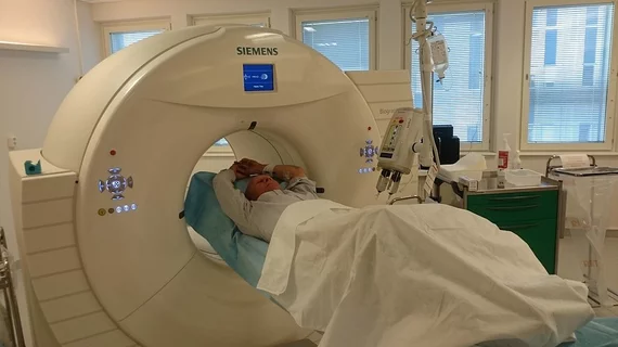
Molecular imaging (also called nuclear medicine or nuclear imaging) can image the function of cells inside the body at the molecular level. This includes the imaging modalities of positron emission computed tomography (PET) and single photon emission computed tomography (SPECT) imaging. How does PET and SPECT imaging work? Small amounts of radioactive material (radiopharmaceuticals) injected into a patient. These can use sugars or chemical traits to bond to specific cells. The radioactive material is taken up by cells that consume the sugars. The radiation emitted from inside the body is detected by photon detectors outside the body. Computers take the data to assemble images of the radiation emissions. Nuclear images may appear fuzzy or ghostly rather than the sharper resolution from MRI and CT.  But, it provides metabolic information at a cellular level, showing if there are defects in the function of the heart, areas of very high metabolic activity associated with cancer cells, or areas of inflammation, data not available from other modalities. These noninvasive imaging exams are used to diagnose cancer, heart disease, Alzheimer’s and Parkinson’s disease, bone disorders and other disorders. 

Largest tau PET study confirms link to memory decline

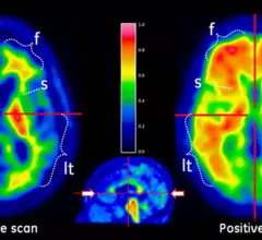
Finding tau deposition in the brain could be a way to verify Alzheimer's prior to the arrival of all the characteristic symptoms, according to a study presented during this week's Alzheimer's Association International Conference (AAIC) 2014.

Thumbnail

AAIC: F-18 Flute shown to change management in 20% of dementia patients

The amyloid PET agent F-18 flutemetamol (Vizamyl) was the topic of two scientific sessions at the Alzheimer’s Association International Conference (AAIC), which concluded yesterday in Copenhagen. Results of one study showed improvements in clinical decision making and clinician confidence as a result of information gleaned from flutemetamol PET. In another study, patients with mild cognitive impairment (MCI) whose scans were positive for flutemetamol were 2.5 times more likely to progress to Alzheimer's disease.

FDA approves gantry for IBA’s small-scale proton system

IBA announced this week that the FDA has provided marketing authorization for the gantry beam line designed for the company’s compact proton therapy system, which will be used much like its larger cousins for the treatment of a variety of cancers.

Thumbnail

Construction of NorthStar’s new radioisotope facility is underway

NorthStar Medical Radioisotopes announced July 16 that the company had broken ground on the 50,000-square-foot production facility and headquarters in Beloit, Wis.

Proton detectors for better therapy

A cutting-edge detector developed by researchers in the United Kingdom and South Africa stands to spark more effective proton therapy for cancer patients.

PET imaging for myocardial innervation expands with F-18 labeled agent

First-in-human study of an investigational radiotracer, F-18 LMI1195, shows clear imaging of myocardial innervation via the norepinephrine transporter system—tipping off potential adverse cardiac events, according to a study published July 3 in the Journal of Nuclear Medicine.

Thumbnail

Dementia numbers falling in some countries and underestimated in others

New cases of dementia are diminished in developed nations, which could be a sign of the successes of preventative care and reduced risk factors. On the other hand, dementia may have been sorely underestimated in countries with lower and middle incomes, according to research presented during the current Alzheimer’s Association International Conference (AAIC) in Copenhagen, Denmark.

AAIC 2014: Alzheimer’s risk factors in sharper relief

A number of factors are standing out as promoters for and protectors against the development of Alzheimer’s disease, according to research presented during the 2014 Alzheimer’s Association International Conference (AAIC) being held July 12-17 at the Bella Center in Copenhagen, Denmark.