Molecular Imaging

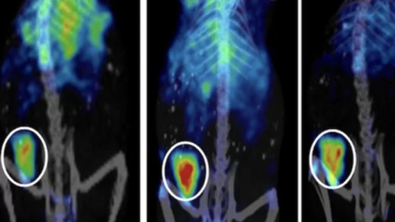
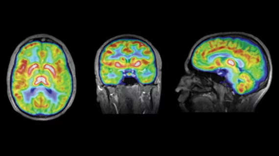
Molecular imaging (also called nuclear medicine or nuclear imaging) can image the function of cells inside the body at the molecular level. This includes the imaging modalities of positron emission computed tomography (PET) and single photon emission computed tomography (SPECT) imaging. How does PET and SPECT imaging work? Small amounts of radioactive material (radiopharmaceuticals) injected into a patient. These can use sugars or chemical traits to bond to specific cells. The radioactive material is taken up by cells that consume the sugars. The radiation emitted from inside the body is detected by photon detectors outside the body. Computers take the data to assemble images of the radiation emissions. Nuclear images may appear fuzzy or ghostly rather than the sharper resolution from MRI and CT.  But, it provides metabolic information at a cellular level, showing if there are defects in the function of the heart, areas of very high metabolic activity associated with cancer cells, or areas of inflammation, data not available from other modalities. These noninvasive imaging exams are used to diagnose cancer, heart disease, Alzheimer’s and Parkinson’s disease, bone disorders and other disorders. 

Thumbnail

FDG PET/CT points out malignant lymph nodes inside the breast

Poor patient outcomes are associated with primary breast cancer that has metastasized to internal mammary lymph nodes. Researchers compared F-18 FDG PET/CT with results of cytopathologic testing from image-guided fine-needle aspiration, revealing a high percentage of proven metastases from positive FDG PET scans, according to a study published in the May issue of the American Journal of Roentgenology.

Spoils of acute myeloid leukemia genetics research revealed

Genomics research peering into the pathogenesis of adult de novo acute myeloid leukemia (AML) reveals many important relationships in both genomic and exomic mutations, according to a review published May 1 in the New England Journal of Medicine.

Navidea Biopharmaceuticals Announces the U.S. Launch of Lymphoseek

Navidea Biopharmaceuticals, Inc. (NYSE MKT:  NAVB), a biopharmaceutical company focused on the development and commercialization of precision diagnostic radiopharmaceuticals, today announced the U.S. launch of Lymphoseek® (technetium Tc 99m tilmanocept) Injection for use in lymphatic mapping procedures that are performed to help in the diagnostic evaluation of potential cancer spread for patients with breast cancer and melanoma.

Alternative neural networks help compensate for Alzheimer's

While verbal memory decline associated with Alzheimer’s disease has been well documented, a similarly relevant area of research has gone relatively uncharted until now. Those affected by the neurodegenerative disease also suffer loss of nonverbal memory function and researchers are mapping the effects of Alzheimer’s disease on brain regions dedicated to this area of cognition and finding newly blazed trails of neural activity, according to a study published April 26 in Neurology.

It adds up: Quantitative PET may help stage lung cancer

For patients with non-small cell lung carcinoma, volumetric data derived from quantitative PET imaging could provide more precision staging and inform physicians about patient survival, according to a study published in the March issue of the American Journal of Roentgenology.

Dual-target PET teases apart dementia class

A PET imaging technique gleaning information about both dopamine terminal integrity and amyloid burden in the brain may help researchers classify forms of mild cognitive impairment (MCI) and predict dementia risk, according to a study published online April 9 in The Journal of Nuclear Medicine.

Education linked w/ cognitive reserve in early-stage Alzheimer’s patients

An increasing number of studies are exploring the possibilities of cognitive reserve in Alzheimer’s patients with higher education levels. Specific neural networks have been found in an area of the prefrontal cortex that light up in educated subjects and compensate for neural damage in the beginning stages of the disease, according to research published in the June issue of The Journal of Nuclear Medicine.

FDG PET/CT outperforms FLT for thyroid cancer imaging

A comparison between glucose mimicking F-18 FDG and F-18 FLT, which tracks cell proliferation, pegged F-18 FDG PET/CT as a better imaging technique for the diagnosis of differentiated thyroid metastases, according to a study published in the April issue of Radiology.