Molecular Imaging

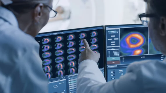
Molecular imaging (also called nuclear medicine or nuclear imaging) can image the function of cells inside the body at the molecular level. This includes the imaging modalities of positron emission computed tomography (PET) and single photon emission computed tomography (SPECT) imaging. How does PET and SPECT imaging work? Small amounts of radioactive material (radiopharmaceuticals) injected into a patient. These can use sugars or chemical traits to bond to specific cells. The radioactive material is taken up by cells that consume the sugars. The radiation emitted from inside the body is detected by photon detectors outside the body. Computers take the data to assemble images of the radiation emissions. Nuclear images may appear fuzzy or ghostly rather than the sharper resolution from MRI and CT.  But, it provides metabolic information at a cellular level, showing if there are defects in the function of the heart, areas of very high metabolic activity associated with cancer cells, or areas of inflammation, data not available from other modalities. These noninvasive imaging exams are used to diagnose cancer, heart disease, Alzheimer’s and Parkinson’s disease, bone disorders and other disorders. 

FDG PET/CT upstages radiography in unmasking multiple myeloma

Although the outlook for patients with multiple myeloma, a cancer that attacks blood plasma and bone marrow, is typically dismal, F-18 FDG PET/CT has emerged as a strategy that could provide earlier diagnosis as well as more accurate treatment monitoring and patient prognosis, according to a review in the April issue of the American Journal of Roentgenology.

Research shows unique dementia profile for those with TBI history

Dementia in people with a history of traumatic brain injury (TBI) may be a clinical phenotype distinct from other known dementia subtypes, according to a study published in the April issue of NeuroRehabilitation.

Cardiac, stroke risk assessments trump dementia scoring as cognitive decline predictors

An evaluation of cardiovascular and stroke risk was found to be a more promising predictor of potential cognitive decline than an assessment used to predict development of dementia, according to a study in the April issue of Neurology.

SPECT/CT signals diabetic bone disease

Hybrid molecular imaging can lead to diagnosis of osteomyelitis in diabetic patients via bedside bone puncture without the need for surgical or image-guided biopsy, according to a study published online in Diabetes Care in March.

Imaging Market: Molecular, Nanoscale, Atomic, Optical, Thermal and Infrared Imaging Industry Analysis in New Research Reports at RnRMarketResearch.com

"Global Markets and Technologies for Molecular, Nanoscale and Atomic Imaging", "Global Thermal Imaging Market 2012-2016", "Global Infrared Imaging Market 2012-2016" and "Optical Imaging Market (2013-2018)" reports are now available with RnR Market Research

CMS proposes to cover PET for most cancers, prostate excluded

The Centers for Medicare & Medicaid Services (CMS) released a proposed decision memo that outlined its suggestion to cover PET imaging for most solid tumors. CMS noted that PET monitoring of Medicare beneficiaries with prostate cancer is not reasonable and necessary.

Future looks bright for thyroid cancer molecular imaging, treatments

A number of research breakthroughs in molecular imaging and molecular-targeted treatments of thyroid cancer are showing signs of expansion into the clinic, according to a review in the March issue of The Lancet.

Amyloid tracer shows diagnostic promise for Alzheimer’s disease

The amyloid imaging agent F-18 florbetaben has been moving forward in clinical trials for the diagnosis of Alzheimer’s disease. The up and coming F-18 florbetaben is showing signs of similar effectiveness, according to research published in the May issue of The Journal of Nuclear Medicine that presented a dynamic model of amyloid imaging using the tracer.