Molecular Imaging

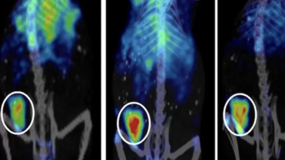
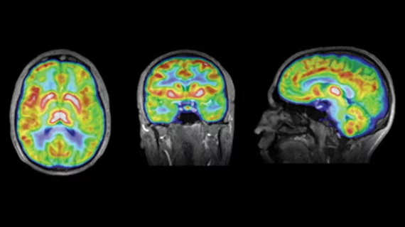
Molecular imaging (also called nuclear medicine or nuclear imaging) can image the function of cells inside the body at the molecular level. This includes the imaging modalities of positron emission computed tomography (PET) and single photon emission computed tomography (SPECT) imaging. How does PET and SPECT imaging work? Small amounts of radioactive material (radiopharmaceuticals) injected into a patient. These can use sugars or chemical traits to bond to specific cells. The radioactive material is taken up by cells that consume the sugars. The radiation emitted from inside the body is detected by photon detectors outside the body. Computers take the data to assemble images of the radiation emissions. Nuclear images may appear fuzzy or ghostly rather than the sharper resolution from MRI and CT.  But, it provides metabolic information at a cellular level, showing if there are defects in the function of the heart, areas of very high metabolic activity associated with cancer cells, or areas of inflammation, data not available from other modalities. These noninvasive imaging exams are used to diagnose cancer, heart disease, Alzheimer’s and Parkinson’s disease, bone disorders and other disorders. 

Value-based Purchasing: What It Means for Performance Evaluation

Now that the fee-for-service reimbursement model may be coming to an end, radiology providers will have to adjust pracice to survive (and succeed) in a value-based environment.

A Merger in Maine

Two large cardiology practices in Maine merged with an integrated delivery network. The integration demanded a complete overhaul of governance, IT considerations and molecular imaging workflow.

Theranostics Dissected

Recent developments in theranostics could usher in an era that helps drive personalized medicine from research to reality.

PET tracer predicts resistance to hormone therapy for breast cancer

18F-labeled fluoromisonidazole (18F-FMISO) PET/CT can be used during breast cancer treatment planning to predict primary endocrine resistance in estrogen receptor (ER)-positive cancers, according to a study published Feb. 11 in the Journal of Nuclear Medicine.

Banner Health secures $16.3M to launch imaging center

The Banner Alzheimer’s Institute is going to build an imaging center for the  research and treatment of Alzheimer’s disease, as well as heart disease and cancer, with $16.3 million in federal grants and donations.

Disparities based on age, race persist in PET use for lung cancer patients

Demographic differences in the use of PET imaging among Medicare beneficiaries with non-small cell lung cancer have persisted since the modality’s approval by Medicare in 1998, according to a study published online Feb. 15 in Radiology.

Two to tango? Do cerebrovascular disease + amyloidosis make for Alzheimer’s?

The combination of white matter hyperintensities and cerebral amyloidosis may result in the clinical manifestation of Alzheimer’s disease (AD), according to a study published Feb. 18 in JAMA Neurology.

Cardiac PET/MR: In pursuit of the killer app

Hybrid PET/MR has demonstrated its potential in cardiac imaging thanks to its superior tissue contrast and ability to deliver multimodal quantitative imaging parameters; however, technical and financial hurdles remain, according to a review article published in the March issue of the Journal of Nuclear Medicine.