Molecular Imaging

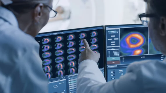
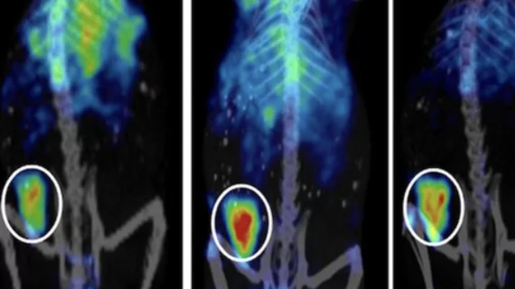
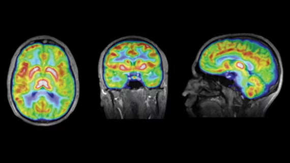
Molecular imaging (also called nuclear medicine or nuclear imaging) can image the function of cells inside the body at the molecular level. This includes the imaging modalities of positron emission computed tomography (PET) and single photon emission computed tomography (SPECT) imaging. How does PET and SPECT imaging work? Small amounts of radioactive material (radiopharmaceuticals) injected into a patient. These can use sugars or chemical traits to bond to specific cells. The radioactive material is taken up by cells that consume the sugars. The radiation emitted from inside the body is detected by photon detectors outside the body. Computers take the data to assemble images of the radiation emissions. Nuclear images may appear fuzzy or ghostly rather than the sharper resolution from MRI and CT.  But, it provides metabolic information at a cellular level, showing if there are defects in the function of the heart, areas of very high metabolic activity associated with cancer cells, or areas of inflammation, data not available from other modalities. These noninvasive imaging exams are used to diagnose cancer, heart disease, Alzheimer’s and Parkinson’s disease, bone disorders and other disorders. 

Quantifying Myocardial Blood Flow: An Expanding Role for PET?

New developments in tracers could make cardiac PET the more precise approach for absolute quantification of myocardial blood flow.

Spotlight On | Breast Imaging

While questions about the efficacy of mammography aboundespecially regarding high-risk women with dense breastsresearch is turning to newer PET-based techniques for more specific diagnosis.

PET/MR: In Search of the Killer App

The final diagnostic form the evolving hybrid PET/MR tree will take is uncertain, but it has already begun to blossom. Thus, the low-hanging clinical fruit has lured a number of medical centers to early adoption. Experts weigh in on the potential that PET/MR may hold for various neurologic, oncologic and cardiovascular conditions.

Is There a Role for PET/MR?

This issue highlights the emergence of PET/MR, which follows the introduction of SPECT/CT and PET/CT that have convincingly shown that multimodality imaging can result in significantly improved disease characterization.

Low-Dose Molecular Breast Imaging: Is It Finally Here?

Gamma Medica

Molecular breast imaging is proving its worth as an low-dose alternative to MRI for women with dense breasts.

18F-FDG PET Breaks New Ground in Small Cell Lung Cancer Imaging

While 18F-FDG PET is the gold standard for evaluating and managing patients with lung cancer, recent data have shown it also could be beneficial in diagnosing, staging and monitoring of small cell lung cancer.

Biomarker Update: Alzheimers Detection

This article discusses the recent updates in Alzheimers detection using amyloid PET radiotracer 18F-flutemetamol, proteins in the cerebrospinal fluid and blood.

Deadline Approaches: How Nuclear Labs Prep for Accreditation

Independent nuclear laboratories are under a mandate to achieve accreditation by Jan. 1, 2012, to continue receiving Medicare reimbursement. While maneuvering the varied accreditation processes requires practice management savvycausing some practices to prolong the processthe impending deadline is approaching fast.