Molecular Imaging

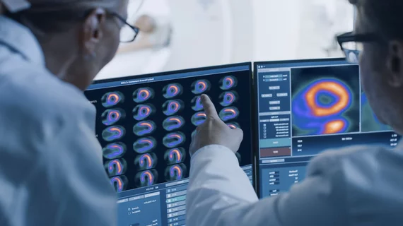
Molecular imaging (also called nuclear medicine or nuclear imaging) can image the function of cells inside the body at the molecular level. This includes the imaging modalities of positron emission computed tomography (PET) and single photon emission computed tomography (SPECT) imaging. How does PET and SPECT imaging work? Small amounts of radioactive material (radiopharmaceuticals) injected into a patient. These can use sugars or chemical traits to bond to specific cells. The radioactive material is taken up by cells that consume the sugars. The radiation emitted from inside the body is detected by photon detectors outside the body. Computers take the data to assemble images of the radiation emissions. Nuclear images may appear fuzzy or ghostly rather than the sharper resolution from MRI and CT.  But, it provides metabolic information at a cellular level, showing if there are defects in the function of the heart, areas of very high metabolic activity associated with cancer cells, or areas of inflammation, data not available from other modalities. These noninvasive imaging exams are used to diagnose cancer, heart disease, Alzheimer’s and Parkinson’s disease, bone disorders and other disorders. 

On the Verge: Inside Clinical Trials that Could Change Patient Care

PET imaging is effective in staging, restaging, detecting recurrence and treatment monitoring across a wide range of cancers.

Electrophysiology Procedure Planning: Using Nuclear Modalities to Help Guide Ablations & Patient Selection

Due to the commonality of these arrhythmias and the number of procedures necessary to treat them, there is a growing need for the field of electrophysiology (EP). And to better treat patients with dangerous arrhythmias, practitioners have begun using cardiac PET and SPECT to help in patient selection and to guide EP procedures including ablations, lead placements and CRT implantations.

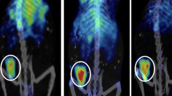
Preclinical to Clinical 18F-FLT PET Imaging Moving from Mice to Men

One of the most promising radiopharmaceuticals is the proliferation marker 3'-deoxy-3'-[18F] fluorothymidine (18F-FLT). 

Monitoring Cancer's Course: Quantitative PET Treatment Response Assessment

FDG-PET is more accurately predicting cancer treatment response than anatomical imaging, allowing more individualized, successful therapeutic strategies and patient management in lymphomas, breast, lung, gastroesophageal, colorectal and many other cancers.

PET/MR: Birth of a New Modality

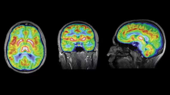
A new imaging modality is coming into clinical focus that promises to improve the diagnosis and monitoring of brain disorders such as Alzheimers, and cancers such as prostate, breast and liver, and even heart disease.

Molecular Breast Imaging: When to Use It

Gamma Medica

There is good news and bad news with breast cancer. While incidence rates have decreased by 2 percent per year between 1999 and 2006 and breast cancer death and diagnosis have largely declined because of improved screening and training techniques, current imaging methods have fallen short on detecting the disease effectively and early in certain patient populations.

SPECT/CT: Defining a Niche in Infection Imaging

SPECT/CT is making notable inroads in imaging infection. It allows the detection of unsuspected infectious foci, especially in areas that are not well investigated by other imaging modalities. It is improving diagnostic performance and treatment management for specific infections such as in bone, the diabetic foot, post-device implantation, fever of unknown origin (FUO), orthopedics and in post transplant patients.

Preclincal Study Digest | Parkinsons, Nanoparticles & Ovarian Cancer

Preclincal Study Digest | Parkinsons, Nanoparticles & Ovarian Cancer