Molecular Imaging

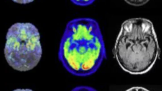
Molecular imaging (also called nuclear medicine or nuclear imaging) can image the function of cells inside the body at the molecular level. This includes the imaging modalities of positron emission computed tomography (PET) and single photon emission computed tomography (SPECT) imaging. How does PET and SPECT imaging work? Small amounts of radioactive material (radiopharmaceuticals) injected into a patient. These can use sugars or chemical traits to bond to specific cells. The radioactive material is taken up by cells that consume the sugars. The radiation emitted from inside the body is detected by photon detectors outside the body. Computers take the data to assemble images of the radiation emissions. Nuclear images may appear fuzzy or ghostly rather than the sharper resolution from MRI and CT.  But, it provides metabolic information at a cellular level, showing if there are defects in the function of the heart, areas of very high metabolic activity associated with cancer cells, or areas of inflammation, data not available from other modalities. These noninvasive imaging exams are used to diagnose cancer, heart disease, Alzheimer’s and Parkinson’s disease, bone disorders and other disorders. 

Preclinical Study Digest

Imaging blood perfusion in pig livers; A molecular theranostics primer; Mice provide a biomarker roadmap for human approach; Simultaneous PET/3D FOT is feasible

ASNC Commits to Patient-Centered Care

The 16th annual scientific session of the American Society of Nuclear Cardiology (ASNC), Sept. 8-11, brings together scientists, clinicians and industry to establish and discuss optimal standards of imaging care.

AJR: A molecular theranostics primer

Molecular theranostics holds promise, offered a review published in the August issue of American Journal of Roentgenology.

The Promise of PET/CT in Oncology

While PET/CT is commonly and very successfully used for the staging and follow-up of cancers, researchers are seeking ways to make the modality more sensitive and specific by using targeted radiotracers and refining scanning techniques.

Inside Cancer Therapy Response and Comparative Effectiveness

PET imaging can play an invaluable role in monitoring the effects of cancer therapies. In my view, cancer therapies that do not achieve early reductions in FDG uptake cannot be effective.

Adding a New Dimension to the Diagnosis & Management of Breast Cancer

A new day is dawning for breast cancer diagnosis, treatment and monitoring with the help of molecular imaging.

Reducing Radiation Dose in Kids: How Low Can You Go

Radiation was brought to the fore within pediatric nuclear medicine following the release of a 2008 study which revealed a chaotic disarray of administered doses within North Americas premier pediatric institutions, including radiopharmaceutical doses varying by factors of as much as 10 in most children and by up to 20 in infants (J Nucl Med 2008; 49:10241027).

How will Comparative Effectiveness Research Impact Molecular Imaging?

Last year, the SNM received a $48,000 grant from the Agency for Healthcare Research and Quality to develop comparative-effectiveness research (CER) of PET and other molecular imaging techniques. The primary emphasis is on the diagnosis and management of cancer patients, but both cardiology and neurology questions are being addressed. Far beyond the dollars, too, is a significant increase in intellectual capital being expended across the globe on the role of CER in molecular imaging.