Molecular Imaging

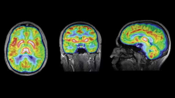
Molecular imaging (also called nuclear medicine or nuclear imaging) can image the function of cells inside the body at the molecular level. This includes the imaging modalities of positron emission computed tomography (PET) and single photon emission computed tomography (SPECT) imaging. How does PET and SPECT imaging work? Small amounts of radioactive material (radiopharmaceuticals) injected into a patient. These can use sugars or chemical traits to bond to specific cells. The radioactive material is taken up by cells that consume the sugars. The radiation emitted from inside the body is detected by photon detectors outside the body. Computers take the data to assemble images of the radiation emissions. Nuclear images may appear fuzzy or ghostly rather than the sharper resolution from MRI and CT.  But, it provides metabolic information at a cellular level, showing if there are defects in the function of the heart, areas of very high metabolic activity associated with cancer cells, or areas of inflammation, data not available from other modalities. These noninvasive imaging exams are used to diagnose cancer, heart disease, Alzheimer’s and Parkinson’s disease, bone disorders and other disorders. 

State of the Heart: PET/CT Imaging

Recently, the value of PET/CT in cardiac imaging has extended beyond the assessment of myocardial viability and perfusion. The combination of PET and CT allows a comprehensive assessment of both structure and function in a single setting and has demonstrated its use in diagnosing and managing coronary artery disease.

Reducing Radiation Dose in Molecular Imaging

Global concern for imaging-based patient radiation dose continues its surge. Recent patient CT radiation overexposure cases in the U.S., studies relating CT scans to higher incidences of cancer and extensive media reporting on radiation exposure have given the issue of radiation exposure during imaging exams an increasingly high profile.

Perspective: Imagings Expanding Role in Stem Cell Therapy

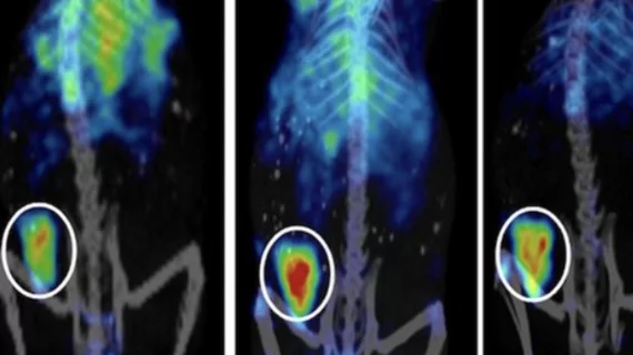
Molecular imaging is playing a key role in direct visualization of stem cells that would allow monitoring of homing and engraftment and survival of transplanted cells in the target tissues, says Antti Saraste, MD, PhD, a cardiologist at Turku PET Centre, Turku University Hospital in Finland.

Movement Disorders: FDOPA PET Guiding Therapy Selection, Moving in to Monitor Drug Therapy

FDOPA PET is showing promise in diagnosing, guiding initial selection of therapy in movement disorders and monitoring functioning of dopamine cell implantation.

New FDA Regulations for the Production of PET Radiopharmaceuticals

Theyve been a long time coming. In December 2009, the FDA released its long-awaited response to the Food and Drug Modernization Act of 1997, signaling a change in the guidelines for PET radiopharmaceutical production which are slated to go into effect on Dec. 12, 2011.

Into the Clinic

This issue of Molecular Imaging Insight addresses various important areas of molecular imaging. The need for reducing radiation dose in molecular imaging is paramount, and is a topic that has gained considerable public attention. Obviously, the ALARA (As Low As Reasonably Achievable) principle applies to PET/CT imaging.

A Clear Path to Reimbursement: Comparative Effectiveness Research

While the recent influx of funding for comparative effectiveness research (CER) from the American Recovery and Reinvestment Act (ARRA) has brought heightened attention to the field in the United States, such well-designed studies have been effectively implemented in Europe, Australia and Asia for the purposes of evaluating molecular imaging technologies and techniques.

Honing in on Lung Cancer

PET/CTsince its introduction in 2001has emerged as the primary diagnostic tool in the staging of lung cancer when compared to PET and CT alone. The shift from CT to PET/CT will continue with technological advances in respiratory gating and molecular imaging.