Molecular Imaging

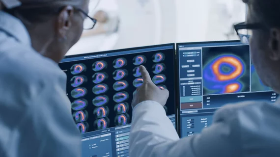
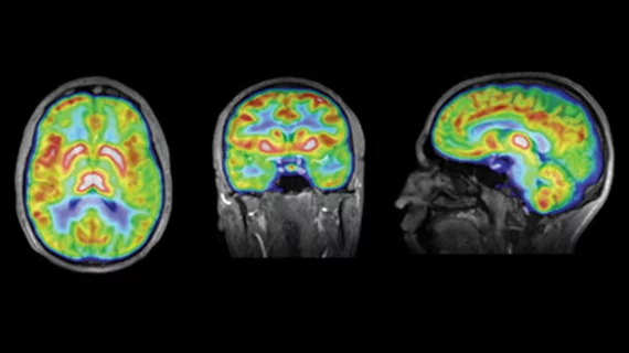
Molecular imaging (also called nuclear medicine or nuclear imaging) can image the function of cells inside the body at the molecular level. This includes the imaging modalities of positron emission computed tomography (PET) and single photon emission computed tomography (SPECT) imaging. How does PET and SPECT imaging work? Small amounts of radioactive material (radiopharmaceuticals) injected into a patient. These can use sugars or chemical traits to bond to specific cells. The radioactive material is taken up by cells that consume the sugars. The radiation emitted from inside the body is detected by photon detectors outside the body. Computers take the data to assemble images of the radiation emissions. Nuclear images may appear fuzzy or ghostly rather than the sharper resolution from MRI and CT.  But, it provides metabolic information at a cellular level, showing if there are defects in the function of the heart, areas of very high metabolic activity associated with cancer cells, or areas of inflammation, data not available from other modalities. These noninvasive imaging exams are used to diagnose cancer, heart disease, Alzheimer’s and Parkinson’s disease, bone disorders and other disorders. 

Hypoxia Imaging in Oncology

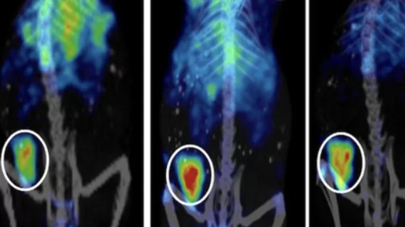
Measuring the oxygen tension, or hypoxia, is critical in determining the effectiveness of radiation therapy, since patients with hypoxic tumors typically have a poor treatment response. Molecular imaging techniques and new imaging biomarkers in development offer a variety of approaches to imaging hypoxic regions in tumorsgoing beyond mere tumor detection to tumor biology characterization for more personalized treatmentintended to improve therapy outcomes and stop the spread of disease.

SPECT/CT's Expansion into Radiology

With the introduction of new molecular imaging modalities, the line between nuclear medicine and radiology blurs a little bit more. While there might have been some tension in the past, with radiologists viewing nuclear medicine as unclear medicine and nuclear medicine physicians operating without the knowledge and experience of cross-sectional imaging, there is now a meeting of the minds of radiologists and nuclear medicine physicians. SPECT/CT utilization is steadily being greeted with more enthusiasm and acceptance within the radiologic community, particularly due to its ability to merge anatomical and functional information, and ultimately, increase diagnostic accuracy and expediency.

Integrated Imaging Comes of Age: Cross-modality Solutions Fuel Better Disease Detection

Truly integrated imaging has arrived. The lines between radiologyand nuclear medicine have blurred with increased utilization ofcross-modality solutions. Several factors are fueling the uptake ofPET/CT—the primary integrated imaging solution.

Peering into the Future: Why We Need New Imaging Biomarkers

The National Cancer Institute and researchers around the globe are exploring and developing an array of new biomarkers that could edge molecular imaging toward routine clinical practice and help deliver on the promise of personalized medicine.

The Role of Imaging Biomarkers

Imaging biomarkers have been developed for use in early cancer diagnosis, staging and restaging of disease and monitoring the effects of therapeutic interventions. In addition, biomarkers for evaluating coronary function and perfusion are well-established. Imaging biomarkers targeting neurodegenerative diseases also are widely used in the clinic.

Getting to the Heart of the Matter: Advanced Coronary Analysis of the Obese Patient

Obesity is a known risk factor for ischemic heart disease, and conducting advanced coronary analysis within this patient population is challenging. SPECT/CT has emerged as a molecular imaging workhorse for evaluating obese patients with coronary artery disease.

Nurturing Connections: From Preclinical Imaging to Diagnosis to Therapy

CLR1404 is entering clinical trials as an anti-cancer therapy agent. It appears well-suited to staging a variety of cancer types and metastatic disease. More specific than FDG, it avoids the complications caused by inflammation or scar tissue. Stay tuned.

Powering the Potential of CT Through Molecular Imaging

In the past decade, CT has established itself as a workhorse in providing anatomical landmarks for molecular imaging. Pairing of the technology with traditional nuclear medicine modalities such as SPECT and PET has propelled the growth of molecular imaging beyond the boundaries of academic research into the mainstream of clinical care.