Molecular Imaging

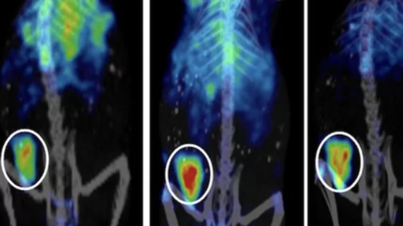
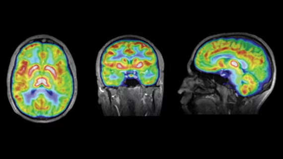
Molecular imaging (also called nuclear medicine or nuclear imaging) can image the function of cells inside the body at the molecular level. This includes the imaging modalities of positron emission computed tomography (PET) and single photon emission computed tomography (SPECT) imaging. How does PET and SPECT imaging work? Small amounts of radioactive material (radiopharmaceuticals) injected into a patient. These can use sugars or chemical traits to bond to specific cells. The radioactive material is taken up by cells that consume the sugars. The radiation emitted from inside the body is detected by photon detectors outside the body. Computers take the data to assemble images of the radiation emissions. Nuclear images may appear fuzzy or ghostly rather than the sharper resolution from MRI and CT.  But, it provides metabolic information at a cellular level, showing if there are defects in the function of the heart, areas of very high metabolic activity associated with cancer cells, or areas of inflammation, data not available from other modalities. These noninvasive imaging exams are used to diagnose cancer, heart disease, Alzheimer’s and Parkinson’s disease, bone disorders and other disorders. 

Diagnostic SPECT/CT: All in Favor

First there was PET imaging, and then emerged the need for anatomic localization. Thus was born the hybrid PET/CT scanner, which currently dominates the market. The acceptance of SPECT/CT is gaining momentum, as more and more clinical uses for the combination imaging are proving advantageous.

Molecular Imaging's Role in Prostate Cancer Imaging

Multimodality molecular imaging and new developments in imaging agents are beginning to make their mark on diagnosis, staging, detecting recurrence and measuring response to therapy in prostate cancerPET/CT with choline is the emerging standard.

Nuclear Medicine & Radiology: A Whole New Integrated World

The development of molecular imaging has not only had a profound impact on medical practice and imaging, but on the relationship between the two established specialties of radiology and nuclear medicine.

A Shift from Conventional Nuclear Medicine to PET

The global healthcare environment is rapidly changing. This is prompted by the overall economic situation and also by the change in healthcare policies and politics that undoubtedly will have a substantial impact on the way medicine is practiced worldwide. 

Industry-academia relationship: The need to collaborate and disclose

The current economic crisis affects industry, but also academia, with decreasing federal funds available for medical research. Many of the brightest researchers create and invent novel approaches in academia. On the other hand, scientific discoveries by pharmaceutical, radiopharmaceutical and instrumentation companies frequently need to be tested and validated in collaboration with academia.

Surviving the Moly shortage: Investing in Alternatives

Over the past few years, the global nuclear medicine community has been plagued with shortages of medical radioisotopes, which is particularly detrimental for the oft-used molybdenum-99 (Mo-99)the parent isotope of technetium-99m (Tc-99m), the most widely utilized radioisotope in the world for molecular and nuclear diagnostic imaging studies. 

A Look Ahead: Inside Optical Imaging

Within the field of molecular imaging, there are quite a few techniques that are well established within routine clinical practice. Other techniques have earned the reputation of being powerful and innovative when it comes to experimental or preclinical research, but have had trouble translating into the clinic. That could be changing, particularly in the area of optical imaging.

PET/MRI: Where We Are, Where Were Going

The merging of PET and MRI is producing synergies far beyond the capabilities of two distinct imaging modalities. The sum is clearly greater than its parts, according to researchers using both preclinical and clinical PET/MRI prototypes, notably because scanning is simultaneous rather than sequential like its PET/CT cousin, and the excellent soft tissue visualization MRI offers. A clinical prototype is being used to study human brain tumors, and researchers predict integrated whole-body human PET/MRI scanners will be available in the next couple of years.