Molecular Imaging

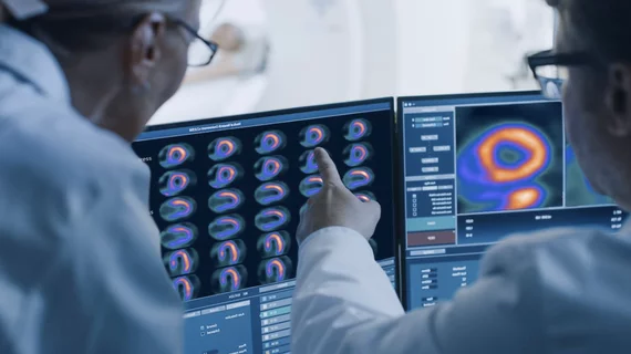
Molecular imaging (also called nuclear medicine or nuclear imaging) can image the function of cells inside the body at the molecular level. This includes the imaging modalities of positron emission computed tomography (PET) and single photon emission computed tomography (SPECT) imaging. How does PET and SPECT imaging work? Small amounts of radioactive material (radiopharmaceuticals) injected into a patient. These can use sugars or chemical traits to bond to specific cells. The radioactive material is taken up by cells that consume the sugars. The radiation emitted from inside the body is detected by photon detectors outside the body. Computers take the data to assemble images of the radiation emissions. Nuclear images may appear fuzzy or ghostly rather than the sharper resolution from MRI and CT.  But, it provides metabolic information at a cellular level, showing if there are defects in the function of the heart, areas of very high metabolic activity associated with cancer cells, or areas of inflammation, data not available from other modalities. These noninvasive imaging exams are used to diagnose cancer, heart disease, Alzheimer’s and Parkinson’s disease, bone disorders and other disorders. 

Preclinical Imaging: The Rapidly Evolving Role of Nanotechnology

Researchers at Memorial Sloan-Kettering Cancer Center, along with collaborators at Cornell University and Hybrid Silica Technologies, have received approval for their first Investigational New Drug Application (IND) from the U.S. Food and Drug Administration (FDA) for an ultrasmall inorganic (silica) nanoparticle platform for tumor targeting and for the treatment of cancers in the future.

Improving Risk Stratification for Cardiovascular Disease

SPECT myocardial perfusion imaging (MPI) is a well-validated noninvasive test to determine if coronary artery disease (CAD) is the cause of a patients chest pain. While SPECT will continue to play a role in this patient population, other tests are helping to fill in the gaps in identifying those at risk of cardiac events.

Proving the Value of Registries

This issue provides updates on several important applications of molecular imaging. The cover story presents novel ideas to better identify patients who are at risk for or have coronary artery disease.

On the Verge: Inside Clinical Trials that Could Change Patient Care

PET imaging is effective in staging, restaging, detecting recurrence and treatment monitoring across a wide range of cancers.

Electrophysiology Procedure Planning: Using Nuclear Modalities to Help Guide Ablations & Patient Selection

Due to the commonality of these arrhythmias and the number of procedures necessary to treat them, there is a growing need for the field of electrophysiology (EP). And to better treat patients with dangerous arrhythmias, practitioners have begun using cardiac PET and SPECT to help in patient selection and to guide EP procedures including ablations, lead placements and CRT implantations.

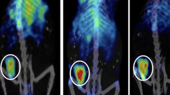
Preclinical to Clinical 18F-FLT PET Imaging Moving from Mice to Men

One of the most promising radiopharmaceuticals is the proliferation marker 3'-deoxy-3'-[18F] fluorothymidine (18F-FLT). 

Monitoring Cancer's Course: Quantitative PET Treatment Response Assessment

FDG-PET is more accurately predicting cancer treatment response than anatomical imaging, allowing more individualized, successful therapeutic strategies and patient management in lymphomas, breast, lung, gastroesophageal, colorectal and many other cancers.

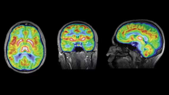
PET/MR: Birth of a New Modality

A new imaging modality is coming into clinical focus that promises to improve the diagnosis and monitoring of brain disorders such as Alzheimers, and cancers such as prostate, breast and liver, and even heart disease.