Molecular Imaging

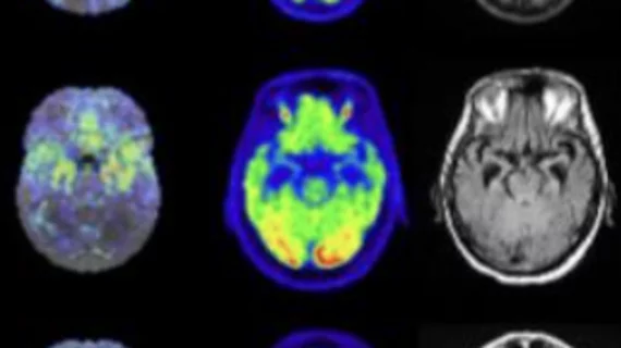
Molecular imaging (also called nuclear medicine or nuclear imaging) can image the function of cells inside the body at the molecular level. This includes the imaging modalities of positron emission computed tomography (PET) and single photon emission computed tomography (SPECT) imaging. How does PET and SPECT imaging work? Small amounts of radioactive material (radiopharmaceuticals) injected into a patient. These can use sugars or chemical traits to bond to specific cells. The radioactive material is taken up by cells that consume the sugars. The radiation emitted from inside the body is detected by photon detectors outside the body. Computers take the data to assemble images of the radiation emissions. Nuclear images may appear fuzzy or ghostly rather than the sharper resolution from MRI and CT.  But, it provides metabolic information at a cellular level, showing if there are defects in the function of the heart, areas of very high metabolic activity associated with cancer cells, or areas of inflammation, data not available from other modalities. These noninvasive imaging exams are used to diagnose cancer, heart disease, Alzheimer’s and Parkinson’s disease, bone disorders and other disorders. 

Molecular Breast Imaging: When to Use It

Gamma Medica

There is good news and bad news with breast cancer. While incidence rates have decreased by 2 percent per year between 1999 and 2006 and breast cancer death and diagnosis have largely declined because of improved screening and training techniques, current imaging methods have fallen short on detecting the disease effectively and early in certain patient populations.

SPECT/CT: Defining a Niche in Infection Imaging

SPECT/CT is making notable inroads in imaging infection. It allows the detection of unsuspected infectious foci, especially in areas that are not well investigated by other imaging modalities. It is improving diagnostic performance and treatment management for specific infections such as in bone, the diabetic foot, post-device implantation, fever of unknown origin (FUO), orthopedics and in post transplant patients.

Preclincal Study Digest | Parkinsons, Nanoparticles & Ovarian Cancer

Preclincal Study Digest | Parkinsons, Nanoparticles & Ovarian Cancer

PET Helps Cushion Nuclear Reimbursement Cuts

Orlando Heart Center, a 23-physician four-office practice close to Orlando, Fla., prepared for the 2010 reimbursement cuts by investing in a PET scanner. The center since has shifted more SPECT patients to PET, resulting in multiple benefits for the practice and patients.

Lessons Learned from the Moly Shortage

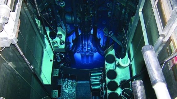
The nuclear medicine community learned many lessons from the shortage of molybdenum-99 (Mo-99), the mother radioisotope of technetium-99m (Tc-99m), during the recent shutdowns of  the Canadian National Research Universal (NRU) reactor in Chalk River, Ontario, and High Flux Reactor (HFR) in Petten, the Netherlands.

Imaging Hearts & Bones PET/CT Underutilization

In this issue, we address several important issues related to the quality of care and the business of molecular imaging. The recent 99mTc shortage made it clear that long established imaging studies will eventually be replaced by more robust and higher quality alternatives. This will foremost affect two major applications of nuclear medicine, myocardial perfusion and bone imaging.

Evidence-based Medicine in Oncology: Molecular Imaging's Expanding Role

Evidence has clearly shown the impact of PET/CT imaging in initial treatment strategiesdiagnosis and initial staging as well as in the subsequent treatment strategiestreatment monitoring and restaging/detection of suspected recurrence in various cancers. The molecular imaging community needs to work with industry and regulators to emphasize that patient-focused care benefits everyone.

Beyond PSA: Prostate Cancer Biomarkers

Prostate cancer is the most common cancer among men worldwide and is the second leading cause of death in men after lung cancer in the U.S.