Molecular Imaging

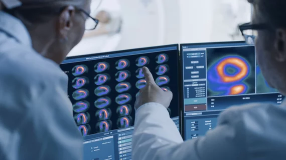
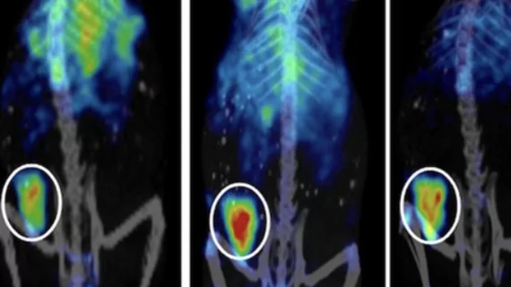
Molecular imaging (also called nuclear medicine or nuclear imaging) can image the function of cells inside the body at the molecular level. This includes the imaging modalities of positron emission computed tomography (PET) and single photon emission computed tomography (SPECT) imaging. How does PET and SPECT imaging work? Small amounts of radioactive material (radiopharmaceuticals) injected into a patient. These can use sugars or chemical traits to bond to specific cells. The radioactive material is taken up by cells that consume the sugars. The radiation emitted from inside the body is detected by photon detectors outside the body. Computers take the data to assemble images of the radiation emissions. Nuclear images may appear fuzzy or ghostly rather than the sharper resolution from MRI and CT.  But, it provides metabolic information at a cellular level, showing if there are defects in the function of the heart, areas of very high metabolic activity associated with cancer cells, or areas of inflammation, data not available from other modalities. These noninvasive imaging exams are used to diagnose cancer, heart disease, Alzheimer’s and Parkinson’s disease, bone disorders and other disorders. 

Pint-size Power: PET/CT and SPECT/CT in Pediatrics

PET/CTa technology whose potential is now being appreciated, but has yet to be fully realizedhas proven successful in the accurate staging and monitoring of oncology patient therapy for a variety of cancers. SPECT/CT, while relatively new, is beginning to make its mark in areas such as oncology, cardiology, neurology imaging. 

Diagnosing Heart Disease

Diagnosing heart disease in women—the No. 1 cause of death of womenacross the globe—is sometimes difficult, but molecular PET and SPECTimaging is beginning to contribute to resolving this problem.

The Ammonia Option: PET/CT Myocardial Perfusion Imaging

13N-ammonia, an established imaging probe for measuringmyocardial perfusion, has now been made available for use in thecommunity. This has thus far been difficult because of the shorthalf-life of 13N-ammonia of about 10 minutes. But even that time factor has been worked out.

Molecular Imaging Training Gaining Traction

As molecular imaging emerges from the cocoon of nuclear medicine, fueled by changes that have made it a vital technique for diagnosing disease and predicting and monitoring treatment response, training for physicians and technologists has made its way to the forefront for many education programs around the United States.

DCE MRI: Standards in the Works

Dynamic contrast agent enhanced MRI (DCE MRI) is a fairly well-established technique that was first introduced in the early 1990s. Used in the pharmaceutical and clinical research environments, the post-processing algorithm evaluates dynamic images to measure contrast agent uptake in target tissue. 

PET/CT Bolsters Sarcoma Armamentarium

Independent of one another, CT and PET have played important roles in the identification and staging of sarcomas. CTs anatomic capabilities have helped physicians locate sarcomas in patients, while PETs functional capabilities have assisted in monitoring treatment, grading sarcoma, separating benign from malignant masses, selecting biopsy sites, and assessing the extent of sarcomas.

Molecular Imagings Next Horizon: Whole-Body SPECT/CT

SPECT is far from the new kid on the molecular imaging block, but except for cardiac imaging, this nuclear medicine modality has not yet realized routine clinical utility as a replacement for conventional planar scanning. Recently developed reconstruction protocols and hybrid SPECT/CT scanners, however, could breathe new life into SPECT by opening the door to clinically feasible whole-body SPECT/CT. The University of Texas M.D. Anderson Cancer Center in Houston, is at the cutting edge of SPECT/CT research. This month, M.D. Anderson experts, Chair of Nuclear Medicine , Associate Professor , and Senior Medical Physicist Bill Erwin from the department of Imaging Physics discuss the clinical path to and potential of whole-body SPECT/CT.

64-slice Cardiac PET/CT: Slice Count Does Matter

Higher quality images and shorter acquisition times than stand-alone PET systems are among the benefits of adding 64-slice CT.