Molecular Imaging

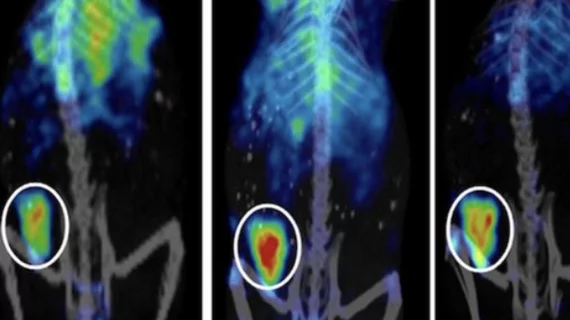
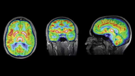
Molecular imaging (also called nuclear medicine or nuclear imaging) can image the function of cells inside the body at the molecular level. This includes the imaging modalities of positron emission computed tomography (PET) and single photon emission computed tomography (SPECT) imaging. How does PET and SPECT imaging work? Small amounts of radioactive material (radiopharmaceuticals) injected into a patient. These can use sugars or chemical traits to bond to specific cells. The radioactive material is taken up by cells that consume the sugars. The radiation emitted from inside the body is detected by photon detectors outside the body. Computers take the data to assemble images of the radiation emissions. Nuclear images may appear fuzzy or ghostly rather than the sharper resolution from MRI and CT.  But, it provides metabolic information at a cellular level, showing if there are defects in the function of the heart, areas of very high metabolic activity associated with cancer cells, or areas of inflammation, data not available from other modalities. These noninvasive imaging exams are used to diagnose cancer, heart disease, Alzheimer’s and Parkinson’s disease, bone disorders and other disorders. 

Expert Panel Defines State of the Art and Future of Molecular Imaging

 A panel of seven clinical experts and public policy leaders offer insight on personalized medicine, technology, biomarkers and the DRA.

Defining Success

PET/CT has come a long way since Time magazine awarded the scanner one of its Medical Inventions of the Year award in December 2000. Published literature since 1996 has established strong evidence for the advantages of PET/CT over PET and CT alone for characterizing lesions as malignant or benign, and for staging, restaging and therapy monitoring of cancer. In addition, emerging applications include imaging of cardiovascular diseases and infection.

Defining Success

PET/CT has come a long way since Time magazine awarded the scanner one of its Medical Inventions of the Year award in December 2000.

Can We Afford NOT to Use FDG-PET?

Numerous clinical studies have compellingly demonstrated that FDG-PET generally outperforms not only individual diagnostic tests, but also a combination of tests to diagnose and stage many cancers. Physicians and patients need to be informed.

DCE MRI: Standards in the Works

Dynamic contrast agent enhanced MRI (DCE MRI) is a fairly well-established technique that was first introduced in the early 1990s. Used in the pharmaceutical and clinical research environments, the post-processing algorithm evaluates dynamic images to measure contrast agent uptake in target tissue. 

To the Bone: SPECT/CT Drives Diagnostic Clarity

Torsten Kuwert, MD, of the University of Erlangen, details how new techniques in SPECT/CT are gaining wider acceptance in diagnosing and staging indeterminate bone lesions—in a single patient visit.

PET Scanning Meets High Definition

Ready to take PET scanning to the next level in its debut this month, high-definition PET (HD•PET) scans optimize image uniformity and enable visualization of smaller structures.

The Big Picture

Molecular imaging is providing new insights into human physiology and disease. It provides more accurate diagnosis, cancer staging, restaging and treatment monitoring, and allows for highly accurate determinations of cardiac and brain function. Molecular imaging thus enables more appropriate and timely treatment decisions.