Molecular Imaging

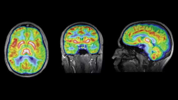
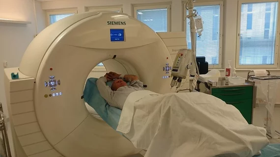
Molecular imaging (also called nuclear medicine or nuclear imaging) can image the function of cells inside the body at the molecular level. This includes the imaging modalities of positron emission computed tomography (PET) and single photon emission computed tomography (SPECT) imaging. How does PET and SPECT imaging work? Small amounts of radioactive material (radiopharmaceuticals) injected into a patient. These can use sugars or chemical traits to bond to specific cells. The radioactive material is taken up by cells that consume the sugars. The radiation emitted from inside the body is detected by photon detectors outside the body. Computers take the data to assemble images of the radiation emissions. Nuclear images may appear fuzzy or ghostly rather than the sharper resolution from MRI and CT.  But, it provides metabolic information at a cellular level, showing if there are defects in the function of the heart, areas of very high metabolic activity associated with cancer cells, or areas of inflammation, data not available from other modalities. These noninvasive imaging exams are used to diagnose cancer, heart disease, Alzheimer’s and Parkinson’s disease, bone disorders and other disorders. 

Non-small Cell Lung Cancer: PET/CT Provides Superior Pre-Radiotherapy Treatment Planning

PET/CT is emerging as the pre-radiotherapy planning imaging study of choice for patients with non-small cell lung cancer (NSCLC), usurping the territory of standalone PET and CT.

Lessons Learned from the Moly Shortage: Is the Crisis Over?

The nuclear medicine community has learned many lessons from the shortage of molybdenum-99 (99Mo), the mother radioisotope of technetium-99m (99mTc) during the recent shut down of both the Canadian National Research Universal (NRU) reactor in Chalk River, Ontario, and High Flux Reactor (HFR) in Petten, the Netherlands. Both these reactors are back online, but are nearing the end of their lifecycle. How are we positioned for the future?

Imaging for Individualized Treatments: Patient Benefits in Oncology, Neurology & Cardiology

As disease treatment becomes more individualized, the spectrum of molecular imaging expands. The role of molecular imaging varies from management of various cancers and differential diagnosis of neurological disorders to identifying patients for cardiac revascularization. Four experts discuss the current and future trends in imaging for individualized treatments in oncology, neurology and cardiology.

Molecular Imaging Predicts Therapeutic Responses Earlier

The earlier we design a treatment and the earlier we know whether it works, the better it is for the patient. Molecular imaging has emerged as a powerful treatment decision-making tool, allowing physicians to make more informed and swift decisions to stop therapy when a patient isnt responding, often very early during the course of treatment. MI is making its mark on a number of cancers, especially breast, esophageal, prostate cancers, gastrointestinal stromal tumor and sarcoma.

Is There a Need for Dedicated PET Imaging Devices?

PET devices have earned dedicated followings in multipurpose imaging in oncology, cardiology and neurology. While PET/CT largely dominates oncology, the demand for PET imaging for specific diseases is increasing, as does the need for low-cost, smaller dedicated scanners in cardiology and neurology.

Radiation Exposure & Hybrid Imaging: Panic Time or Not?

The issue of radiation dose is certainly an attention-grabber these daysnot only within the medical community, but from the popular press as well. And the U.S. government also is enhancing its profile in this area, as evidenced by a Congressional hearing held on medical radiation in February, as well as an announcement by the FDA (accompanied by a two-day public hearing on the subject) that it will be working with manufacturers of advanced medical imaging devices to take steps to prevent patients from receiving excessive doses of radiation. So is there a hint of radiation exposure panic in the air in hybrid imaging?

Imaging Diabetics in the U.S.: Cost Remains PETs Final Barrier

While the diabetic population continues to grow rapidly across the U.S., SPECT holds strong as the gold standard for imaging diabetics, despite the improved prognostic value of PET especially for more complex disease states. Due to the unstable reimbursement environment in the U.S. and large upfront costs of PET, nuclear cardiologists are forced to weigh their options to provide the best patient care, while remaining financially viable.  

How Much Is Enough? A Global Perspective On Technologist Training

The introduction of hybrid imaging, particularly PET/CT, has succeeded in changing the practice of nuclear medicine imaging. Joining CT with PET has lead to calls in various corners of the globe to reappraise who should operate hybrid PET/CT units and what kind of education and training are necessary for nuclear medicine and radiologic technologists to assure safety and effectiveness of each aspect of the study.