Molecular Imaging

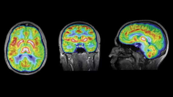
Molecular imaging (also called nuclear medicine or nuclear imaging) can image the function of cells inside the body at the molecular level. This includes the imaging modalities of positron emission computed tomography (PET) and single photon emission computed tomography (SPECT) imaging. How does PET and SPECT imaging work? Small amounts of radioactive material (radiopharmaceuticals) injected into a patient. These can use sugars or chemical traits to bond to specific cells. The radioactive material is taken up by cells that consume the sugars. The radiation emitted from inside the body is detected by photon detectors outside the body. Computers take the data to assemble images of the radiation emissions. Nuclear images may appear fuzzy or ghostly rather than the sharper resolution from MRI and CT.  But, it provides metabolic information at a cellular level, showing if there are defects in the function of the heart, areas of very high metabolic activity associated with cancer cells, or areas of inflammation, data not available from other modalities. These noninvasive imaging exams are used to diagnose cancer, heart disease, Alzheimer’s and Parkinson’s disease, bone disorders and other disorders. 

Automated analysis of cardiac SPECT on par with experts

Fully automated quantitative review of attenuation-corrected and noncorrected myocardial perfusion SPECT data provided performance at least equivalent to expert analysis for detection of stenosis of 70 percent or greater, according to a study published online Jan. 11 in The Journal of Nuclear Medicine.

Additional imaging often recommended, seldom pursued, in PET/CT reports

Nuclear medicine physicians and radiologists recommended additional imaging in approximately one-third of PET/CT reports, but more than half of these recommendations were unnecessary, according to a study published in the January issue of Clinical Radiology. Ordering clinicians followed recommendations for further imaging in less than one-third of the reports.

Fluorescence imaging could help surgeons pinpoint metastatic lymph nodes

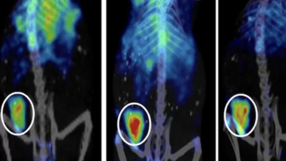
A molecular-targeted imaging method has depicted lymph node metastases intraoperatively in mice. In addition to providing real-time, accurate information about lymph node status, this pathology technique could set the stage for shorter operating room time and reduce unnecessary removal of healthy lymph nodes, according to the researchers whose findings will be published Jan. 15 in Cancer Research.

Reining in Costs of Low-risk CAD Evals

Tests such as SPECT, PET and MRI offer noninvasive alternatives for diagnosing patients with suspected coronary artery disease (CAD). Some recent evidence tilts in favor of these advanced imaging modalities, particularly when a multistep strategy is applied to diagnose CAD. By ruling out low-risk patients, these screening tools may help to eliminate unnecessary treatments and their associated costs.

New Frontiers: Molecular Imaging & the OR

Molecular imaging is spurring dramatic shifts in medicine. The latest venue to witness the revolution may be the surgical suite. The Advanced Multimodality Image-Guided Operating (AMIGO) suite at Brigham and Women’s Hospital (BWH) at Harvard Medical School in Boston weds intraoperative imaging and navigation systems in a surgical environment to set the stage for intraoperative applications of molecular imaging.

Evaluating the Evidence SPECT/CT & Thyroid Cancer

The capability to obtain complementary functional and anatomic information through a single device during a single session makes SPECT/CT an attractive option for numerous applications. In recent years, researchers have shown that SPECT/CT significantly improved the interpretation of planar studies of patients with thyroid cancer in the post-therapy setting. SPECT/CT is now being incorporated in the diagnostic setting to help in the post-operative staging, risk stratification and management of thyroid cancer patients. But is the evidence sufficient to change practice?

Molecular Imaging Merges Care & Cure

Sponsored by Philips Healthcare

A partnership works best when it enhances and rewards its partners. That is the case with the partnership between University Hospitals (UH) Case Medical Center and Case Western Reserve University in Cleveland and Philips Healthcare that is focused on developing, advancing and enhancing medical and molecular imaging technology. It also benefits another very important group—patients.

Less is Not Always Better

This issue of Molecular Imaging Insight focuses on test accuracy, cost and outcomes—all very key topics in 2012.