Molecular Imaging

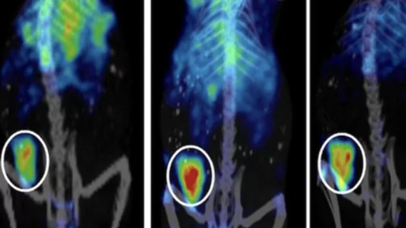
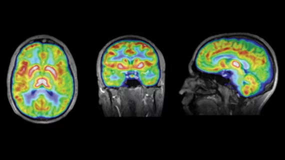
Molecular imaging (also called nuclear medicine or nuclear imaging) can image the function of cells inside the body at the molecular level. This includes the imaging modalities of positron emission computed tomography (PET) and single photon emission computed tomography (SPECT) imaging. How does PET and SPECT imaging work? Small amounts of radioactive material (radiopharmaceuticals) injected into a patient. These can use sugars or chemical traits to bond to specific cells. The radioactive material is taken up by cells that consume the sugars. The radiation emitted from inside the body is detected by photon detectors outside the body. Computers take the data to assemble images of the radiation emissions. Nuclear images may appear fuzzy or ghostly rather than the sharper resolution from MRI and CT.  But, it provides metabolic information at a cellular level, showing if there are defects in the function of the heart, areas of very high metabolic activity associated with cancer cells, or areas of inflammation, data not available from other modalities. These noninvasive imaging exams are used to diagnose cancer, heart disease, Alzheimer’s and Parkinson’s disease, bone disorders and other disorders. 

Advanced MRI offers detailed info on brain lesions in mTBI patients

MRI soon after a mild concussion, or mild traumatic brain injury (mTBI), can detect small lesions and may help refine treatment for people with mTBI, according to research that will be presented at the American Academy of Neurology annual meeting in San Diego, March 16 to 23.

Lantheus Medical Imaging taps Orlandi as next CMO

Lantheus Medical Imaging has appointed Cesare Orlandi, MD, to serve as its next chief medical officer (CMO).

DEX sedative is safe, effective for pediatric imaging patients

Dexmedetomidine (DEX), approved in 2008 by the FDA for use in sedation, may offer a safe, effective and efficient alternative to sedatives currently used in pediatric nuclear medicine imaging, according to a study published in the February edition of Radiology. DEX presents itself as a safer method of sedation during a time of growing demand for pediatric PET oncologic and epileptic scanning.

DTI data ties cognitive impairment in MS patients to signs of brain damage

A diffusion tensor imaging (DTI) study has shown that multiple sclerosis (MS) patients with cognitive problems have more damage to areas of the brain involved with cognitive processes compared with MS patients who don’t suffer from cognitive problems, according to the results of the study published online March 6 in Neurology.

Promise & Downfall of Personalized Medicine

I have been talking to various audiences over the past five years about the potential of molecular imaging in the care of our patients.

PET Could Outshine Other Modalities for CAD

While there has been a movement toward the advocacy of the use of PET imaging for the detection of coronary artery disease (CAD) due to higher resolution and new promising tracers, but whether the modality will trump CT or SPECT as the gold standards remains to be seen.

Tipping the Scales: Molecular Imaging & The Obese Patient

The ripple effect of the obesity epidemic is felt through the entire healthcare system, and for molecular imaging, it affects safety, efficiency and diagnostic quality.

Florbetapir: Alzheimer’s Imaging Inches Into Practice

The practice management considerations, as well as uncertainty about the meaning of a beta-amyloid, scan are impacting early adopters.