Molecular Imaging

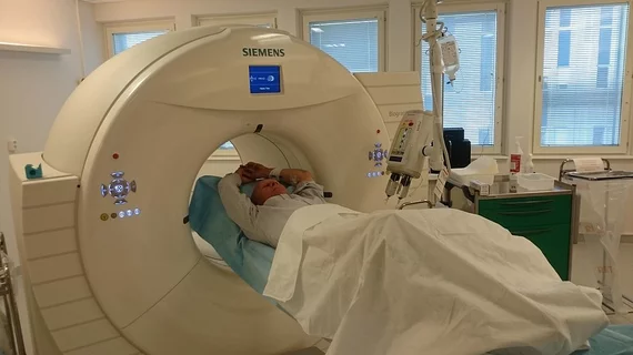
Molecular imaging (also called nuclear medicine or nuclear imaging) can image the function of cells inside the body at the molecular level. This includes the imaging modalities of positron emission computed tomography (PET) and single photon emission computed tomography (SPECT) imaging. How does PET and SPECT imaging work? Small amounts of radioactive material (radiopharmaceuticals) injected into a patient. These can use sugars or chemical traits to bond to specific cells. The radioactive material is taken up by cells that consume the sugars. The radiation emitted from inside the body is detected by photon detectors outside the body. Computers take the data to assemble images of the radiation emissions. Nuclear images may appear fuzzy or ghostly rather than the sharper resolution from MRI and CT.  But, it provides metabolic information at a cellular level, showing if there are defects in the function of the heart, areas of very high metabolic activity associated with cancer cells, or areas of inflammation, data not available from other modalities. These noninvasive imaging exams are used to diagnose cancer, heart disease, Alzheimer’s and Parkinson’s disease, bone disorders and other disorders. 

Radiation Therapy leaders gather for ProteusONE demonstration

IBA (Ion Beam Applications S.A.), the global high-tech leader in next generation radiation therapy and diagnostics for the treatment of cancer, is pleased to announce that today 60 radiation therapy leaders travelled to Belgium to discover the first Proteus®ONE in factory.

Do the math: Best practices for pulmonary nodule management

Out of a range of mathematical methods for managing pulmonary nodules with FDG PET/CT, one quantitative CT technique takes the lead--dynamic area-detector CT analyzed using the dual-input maximum slope method, according to a study published May 22 in the American Journal of Roentgenology.

Advanced SPECT and stress-only MPI lower occupational dose

Many studies have maintained that the most advanced SPECT cameras can significantly reduce patient dose, but what about medical staff? A new protocol of myocardial perfusion SPECT imaging combines state-of-the-art detector technology, image reconstruction and stress-only imaging, all precipitating an up to 60 percent drop in radiation exposure to healthcare workers, according to a study published May 30 in the Journal of Nuclear Medicine.

Patients and Technologists Benefit from Reduced Dose of UltraSPECT Image Reconstruction Software

UltraSPECT, a leading provider of nuclear medicine (NM) image reconstruction technology that reduces radiopharmaceutical dose and acquisition time, announces today the installation of its proprietary Wide Beam Reconstruction (WBR™) software at nearly ten healthcare facilities.

Report predicts continued boom in molecular imaging

Molecular imaging has been in a growth phase since 2010, spurred by emerging markets and demand for hybrid and other cutting edge scanner and biomarker technologies, according to a market report profiling the most recent developments in molecular imaging published in April by UK-based Espicom Business Intelligence.

Cleveland researchers launch Amyvid study

A team of radiologists and neurologists at University Hospitals Case Medical Center in Cleveland are enrolling patients with suspected early-stage Alzheimer’s disease in a study designed to determine if Amyvid can identify amyloid plaques via PET or MRI. Read more about the research by clicking the link below.

Healthcare reform launches new era for nuclear medicine

While healthcare reform has been slowly evolving as a result of many acts of Congress during the past decade, it is the Affordable Care Act of 2010 (ACA), that is driving major changes in healthcare coverage, the economics of the medical industry and quality of care. The latter is being carried by a series of reporting and incentive programs that have a direct impact on the practice of nuclear medicine and molecular imaging, according to a review published March 1 in the Journal of Nuclear Medicine Technology.

Dynamic susceptibility contrast MRI could tip off early-stage Alzheimer’s

Cerebral blood flow (CBF) is an emerging indication of Alzheimer’s pathology and dynamic susceptibility contrast (DSC) MRI may be an ideal modality to track CBF in high-risk patients with mild cognitive impairment, as well as those already diagnosed with the disease, according to a study published in the June issue of Academic Radiology.