Molecular Imaging

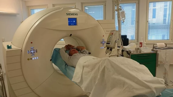
Molecular imaging (also called nuclear medicine or nuclear imaging) can image the function of cells inside the body at the molecular level. This includes the imaging modalities of positron emission computed tomography (PET) and single photon emission computed tomography (SPECT) imaging. How does PET and SPECT imaging work? Small amounts of radioactive material (radiopharmaceuticals) injected into a patient. These can use sugars or chemical traits to bond to specific cells. The radioactive material is taken up by cells that consume the sugars. The radiation emitted from inside the body is detected by photon detectors outside the body. Computers take the data to assemble images of the radiation emissions. Nuclear images may appear fuzzy or ghostly rather than the sharper resolution from MRI and CT.  But, it provides metabolic information at a cellular level, showing if there are defects in the function of the heart, areas of very high metabolic activity associated with cancer cells, or areas of inflammation, data not available from other modalities. These noninvasive imaging exams are used to diagnose cancer, heart disease, Alzheimer’s and Parkinson’s disease, bone disorders and other disorders. 

Epilepsy imaging: Move over C-11 for fluorine-based FMZ PET

C-11 flumazenil (FMZ) PET has been found to have superior localizing specificity compared with F-18 FDG PET for patients with refractory focal epilepsy. However, there are logistical limitations inherent in the use of C-11. A promising potential alternative is F-18 FMZ, according to a study published July 15 in the Journal of Nuclear Medicine.

RadNet Joins Partnership to Explore Potential Alzheimer's Disease Drug

RadNet Inc. (Nasdaq:RDNT), a national leader in providing high-quality, cost-effective, fixed-site outpatient diagnostic imaging services through a network of 248 subsidiary-owned and/or operated outpatient imaging centers, announced its participation in a clinical trial to evaluate the biomarker effects of the investigational drug E2609, a BACE inhibitor, which is being developed for the potential disease modifying treatment of Alzheimer's disease (AD).

Adrenal nodule PET/CT: Qualitative + quantitative eval—either/or or both?

Evaluation of adrenal nodules with F-18 FDG PET/CT was given a mean score of 93 percent diagnostic accuracy with an almost equally high percentage of interreader agreement, suggesting subjective visual interpretation is an effective method of evaluation, according to a review published in the August issue of Academic Radiology.

Thumbnail

Subtyping improves selection for breast cancer patients facing chemotherapy

Adjuvant chemotherapy may not be the best option for patients with certain subtypes of breast cancer who show no clear benefit, according to an extensive retrospective of four clinical trials published June 12 in Breast Cancer Research and Treatment.

Optimizing dose for Lu-177 DOTA-rituximab lymphoma therapy

Radioimmunotherapy with Lu-177 DOTA-rituximab represents a novel and well-matched treatment for B-cell Lymphoma, but no consensus on standardized dosimetry was available, until now, according to research published in the July issue of Journal of Nuclear Medicine.

Edging closer to PET monitoring of therapy response in Hodgkin lymphoma

Personalized therapy assessment for Hodgkin lymphoma (HL) with interim FDG PET could help step up or step down treatment to find the curative sweet spot, but the transition from PET-adaptive clinical trials to practice requires additional time to prove broad benefit for patients, according to a review published July 1 in the Journal of Nuclear Medicine.

CMS releases amyloid PET draft decision

The Centers for Medicare & Medicaid Services (CMS) has released its draft decision for coverage of amyloid PET imaging for the evaluation of dementia and neurodegenerative diseases, including Alzheimer’s. CMS is proposing coverage with evidence development (CED) as of the July 3 memo, which allows one scan to rule out Alzheimer’s pathology in the brain for patients in eligible clinical trials and it would require clinicians to provide detailed data about amyloid PET imaging to gather a body of evidence to make the case for reimbursement of procedures in this category.

Can dedicated cardiac SPECT outperform traditional gamma imaging?

For patients who have just suffered myocardial infarction due to CAD and heart failure, gleaning information about left ventricular (LV) viability with gated blood-pool imaging is highly recommended. Dedicated cardiac SPECT cameras with state-of-the-art detectors and image processing offer a new alternative that may provide advantages compared with traditional SPECT systems.