Molecular Imaging

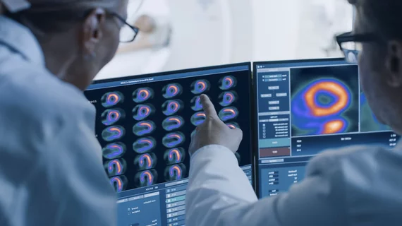
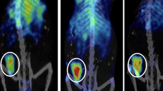
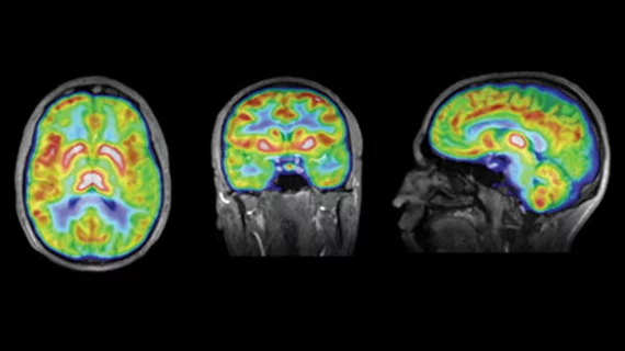
Molecular imaging (also called nuclear medicine or nuclear imaging) can image the function of cells inside the body at the molecular level. This includes the imaging modalities of positron emission computed tomography (PET) and single photon emission computed tomography (SPECT) imaging. How does PET and SPECT imaging work? Small amounts of radioactive material (radiopharmaceuticals) injected into a patient. These can use sugars or chemical traits to bond to specific cells. The radioactive material is taken up by cells that consume the sugars. The radiation emitted from inside the body is detected by photon detectors outside the body. Computers take the data to assemble images of the radiation emissions. Nuclear images may appear fuzzy or ghostly rather than the sharper resolution from MRI and CT.  But, it provides metabolic information at a cellular level, showing if there are defects in the function of the heart, areas of very high metabolic activity associated with cancer cells, or areas of inflammation, data not available from other modalities. These noninvasive imaging exams are used to diagnose cancer, heart disease, Alzheimer’s and Parkinson’s disease, bone disorders and other disorders. 

Edging closer to PET monitoring of therapy response in Hodgkin lymphoma

Personalized therapy assessment for Hodgkin lymphoma (HL) with interim FDG PET could help step up or step down treatment to find the curative sweet spot, but the transition from PET-adaptive clinical trials to practice requires additional time to prove broad benefit for patients, according to a review published July 1 in the Journal of Nuclear Medicine.

CMS releases amyloid PET draft decision

The Centers for Medicare & Medicaid Services (CMS) has released its draft decision for coverage of amyloid PET imaging for the evaluation of dementia and neurodegenerative diseases, including Alzheimer’s. CMS is proposing coverage with evidence development (CED) as of the July 3 memo, which allows one scan to rule out Alzheimer’s pathology in the brain for patients in eligible clinical trials and it would require clinicians to provide detailed data about amyloid PET imaging to gather a body of evidence to make the case for reimbursement of procedures in this category.

Can dedicated cardiac SPECT outperform traditional gamma imaging?

For patients who have just suffered myocardial infarction due to CAD and heart failure, gleaning information about left ventricular (LV) viability with gated blood-pool imaging is highly recommended. Dedicated cardiac SPECT cameras with state-of-the-art detectors and image processing offer a new alternative that may provide advantages compared with traditional SPECT systems. 

Fused PET and MR make the grade in pediatric brain cancers

A connection between cerebral blood volume (CBV) and tumor metabolism in 3D PET and MR characterized a range of pediatric brain tumors. The two modalities were found to sync complementary functional and metabolic information in 94 percent of cases and could have an impact on risk stratification and prognosis with further study, according to research published June 25 in the Journal of Nuclear Medicine.

Phantom fluids: Finding a solution for PET/MR

Steady advancements in simultaneous PET/MR technology make the need for new phantom designs more salient, but the challenge is tricky due to the physics involved in MR imaging. While PET research usually involves watery models, glycols in combination with precise adjustments in tracer composition may be the ideal solution, according to a study published June 21 in the Journal of Nuclear Medicine.

Quantitative PET/CT could improve prostate cancer treatment

Men suffering from prostate cancer have a very high chance of developing bone metastases, making it imperative to track and effectively treat local and distant tumors, especially in castrate-resistant cases. Metabolic imaging with FDG PET could provide a biomarker for predicting patients’ survival, which has been found to be a beneficial indicator in the prognosis of patients with this highly variable disease, according to a study published June 19 in the Journal of Nuclear Medicine.

MR maps tumor angiogenesis with targeted nanoparticles

Effective imaging of the formation of new blood vessels feeding invasive tumors is essential to the continued development of antiangiogenetic therapy. A range of imaging methods and antiangiogenic drugs have been developed in this area with inconsistent results, but a technique using 3D MR and avb3-receptor targeted nanoparticles is providing an index of new vessel development over time that could push research forward and potentially improve patient management and outcomes of antiangiogenetic therapy, according to a study published online June 14 in Radiology.

Experts preach caution after CMS decision for PET cancer imaging

CMS effectively ushered in a fresh start for oncologic PET imaging upon the final coverage decision for PET imaging of solid tumors announced on June 11. Elaboration of new coverage was provided on June 13 and the NOPR (the National Oncologic Pet Registry) was concluded as of June 11. Representatives from the Society of Nuclear Medicine and Molecular Imaging and NOPR moderated a webinar June 19 to discuss the decision and to counsel clinicians on how PET imaging is expected to move forward.