Molecular Imaging

Molecular imaging (also called nuclear medicine or nuclear imaging) can image the function of cells inside the body at the molecular level. This includes the imaging modalities of positron emission computed tomography (PET) and single photon emission computed tomography (SPECT) imaging. How does PET and SPECT imaging work? Small amounts of radioactive material (radiopharmaceuticals) injected into a patient. These can use sugars or chemical traits to bond to specific cells. The radioactive material is taken up by cells that consume the sugars. The radiation emitted from inside the body is detected by photon detectors outside the body. Computers take the data to assemble images of the radiation emissions. Nuclear images may appear fuzzy or ghostly rather than the sharper resolution from MRI and CT.  But, it provides metabolic information at a cellular level, showing if there are defects in the function of the heart, areas of very high metabolic activity associated with cancer cells, or areas of inflammation, data not available from other modalities. These noninvasive imaging exams are used to diagnose cancer, heart disease, Alzheimer’s and Parkinson’s disease, bone disorders and other disorders. 

Adenosine SPECT imaging could single out neurodegenerative, psychiatric disorders

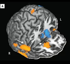
The neuromodulator adenosine and its interaction with a receptor known as A2A are being implicated in a range of brain diseases, including Parkinson’s and Huntington’s, as well as a spate of psychiatric disorders. A novel SPECT agent has been found to successfully shine a spotlight on this relationship, according to research published Aug. 22 in the Journal of Nuclear Medicine.

Exploring the M.O. of drug-resistant cancers

Researchers are forever seeking to outsmart therapy-resistant cancers. "No Through Road," a feature published Aug. 5 in Cancer Today, a publication of the American Association for Cancer Research, highlights just this quest.

Siemens Healthcare introduces continuous FlowMotion PET-CT

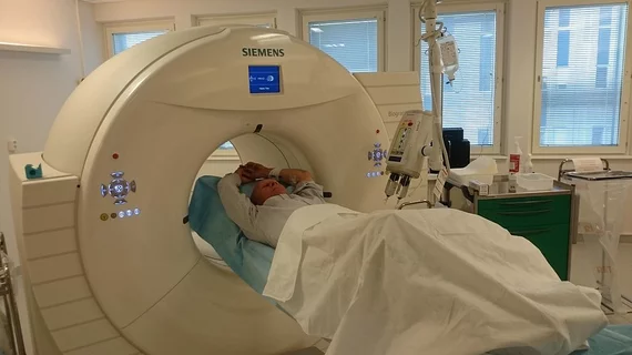
Siemens Healthcare has launched its new Biograph mCT Flow – a groundbreaking PET-CT system. For the first time ever the system overcomes the limitations of conventional bed-based PET-CT with FlowMotion, an innovative new technology that moves the patient smoothly through the system’s gantry, while continuously acquiring PET data.

Navidea awarded NIH SBIR grant for NAV4694 beta-amyloid imaging agent phase III clinical program aimed at Alzheimer’s disease

Navidea Biopharmaceuticals announces award of a Small Business Innovation Research (SBIR) grant from the National Institute On Aging (NIA) of the National Institutes of Health (NIH) in connection with the Company's Phase 3 clinical program for its NAV4694 beta-amyloid imaging agent as an aid in the differential diagnosis of Alzheimer’s disease.

Navidea Biopharmaceuticals signs manufacturing agreement with Siemens’ PETNET Solutions for NAV4694 beta-amyloid imaging agent

Navidea has signed an agreement with Siemens’ PETNET Solutions that grants PETNET Solutions the right to manufacture Navidea’s Fluorine-18 labeled NAV4694, an investigational beta-amyloid PET imaging agent, which is currently being evaluated in Phase 2 and 3 clinical trials evaluating subjects with signs or symptoms of cognitive impairment such as Mild Cognitive Impairment and Alzheimer’s disease.

Regadenoson stress PET/CT MPI trumps SPECT for CAD detection

Regadenoson stress Rb-82 PET/CT myocardial perfusion imaging (MPI) is highly sensitive, 92 percent, for the detection of obstructive CAD and beats SPECT MPI, according to a study published online Aug. 12 in the Journal of Nuclear Medicine.

Atomic imaging reveals how flame retardants compete with estrogens in the body

Flame retardants mimic estradiols in the body, potentially causing havoc on natural endocrine homeostasis, according to new 3D x-ray crystallography research published online Aug. 19 in Environmental Health Perspectives.

Novel oncologic PET tracer captures cell death

A new facet of cancer imaging targets apoptosis, the process of cell death, to help select and monitor anticancer therapies, according to a study published Aug. 15 in the Journal of Nuclear Medicine.