Molecular Imaging

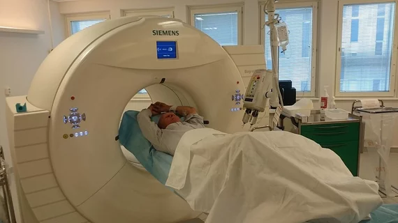
Molecular imaging (also called nuclear medicine or nuclear imaging) can image the function of cells inside the body at the molecular level. This includes the imaging modalities of positron emission computed tomography (PET) and single photon emission computed tomography (SPECT) imaging. How does PET and SPECT imaging work? Small amounts of radioactive material (radiopharmaceuticals) injected into a patient. These can use sugars or chemical traits to bond to specific cells. The radioactive material is taken up by cells that consume the sugars. The radiation emitted from inside the body is detected by photon detectors outside the body. Computers take the data to assemble images of the radiation emissions. Nuclear images may appear fuzzy or ghostly rather than the sharper resolution from MRI and CT.  But, it provides metabolic information at a cellular level, showing if there are defects in the function of the heart, areas of very high metabolic activity associated with cancer cells, or areas of inflammation, data not available from other modalities. These noninvasive imaging exams are used to diagnose cancer, heart disease, Alzheimer’s and Parkinson’s disease, bone disorders and other disorders. 

Siemens’ PETNET Solutions to provide PET radiopharmaceutical services for the US Oncology Network

Siemens’ PETNET Solutions has announced a three-year agreement with The US Oncology Network to supply its entire portfolio of Food and Drug Administration (FDA)-approved PET radiopharmaceutical agents at each of PETNET Solutions’ current Good Manufacturing Practices (cGMP)-certified locations throughout the United States.

Thumbnail

Double duty: Cancer therapy moonlights as breast tumor imaging agent

Metastatic breast cancers positive for HER2 receptor expression can be effectively imaged with Cu-64 DOTA-trastuzumab, traditionally used as a therapeutic drug, according to a study published online Sept. 12 in the Journal of Nuclear Medicine.

WMIC brings the world of molecular imaging to Georgia this week

The World Molecular Imaging Congress being held in Savannah, Ga., from September 18 – 21 is in full swing and bringing new focus to the worldwide molecular imaging agenda.

Definiens and Advanced Cell Diagnostics launch software for quantitative RNA in situ hybridization

Definiens AG, a healthcare company that advances personalized medicine through image analysis and digital pathology solutions, and Advanced Cell Diagnostics (ACD) of Hayward California, a leader in molecular pathology, announced recently the commercial launch of RNAscope® SpotStudio™, a custom-designed image analysis software application for ACD's RNAscope®Assays to detect and quantify RNA biomarkers.

VCAM-1 imaging in ovarian cancer may improve survival

Homing in on vascular cell adhesion molecule-1 (VCAM-1) expression in metastatic ovarian cancer with SPECT could gauge response to therapy and potentially improve survival, according to a study published Sept. 12 in the Journal of Nuclear Medicine.

Technetium kit shortage until mid-October

Jubilant DraxImage, a distributor of the MAA lyophilized Technetium-99m kit, announced that as of Sept. 6 a disruption in the company’s supply chain would effectively render the technetium-synthesis kit out-of-stock until an unspecified date in October, according to a letter to customers.

PET/CT: 32% dose reduction possible with CT optimization

Standardized CT protocols can lead to a substantial decline in radiation dose, according to a large-scale study published in the August issue of American Journal of Roentgenology.

ASNC 2013 in Chicago ensures that cardiologists don’t skip a beat

The 18th annual scientific session of the American Society of Nuclear Cardiology will provide a finger on the pulse of cardiac molecular imaging and medicine later this month at the Hyatt Regency in Chicago from Sept. 26 – 29.