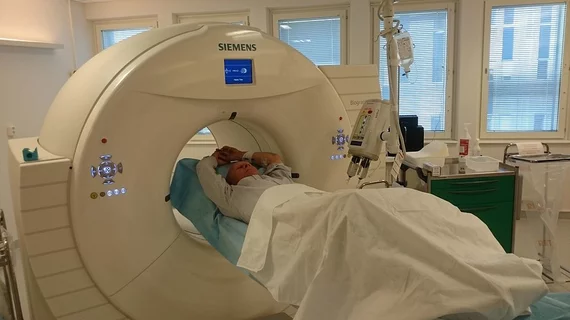
Molecular Imaging

Molecular imaging (also called nuclear medicine or nuclear imaging) can image the function of cells inside the body at the molecular level. This includes the imaging modalities of positron emission computed tomography (PET) and single photon emission computed tomography (SPECT) imaging. How does PET and SPECT imaging work? Small amounts of radioactive material (radiopharmaceuticals) injected into a patient. These can use sugars or chemical traits to bond to specific cells. The radioactive material is taken up by cells that consume the sugars. The radiation emitted from inside the body is detected by photon detectors outside the body. Computers take the data to assemble images of the radiation emissions. Nuclear images may appear fuzzy or ghostly rather than the sharper resolution from MRI and CT.  But, it provides metabolic information at a cellular level, showing if there are defects in the function of the heart, areas of very high metabolic activity associated with cancer cells, or areas of inflammation, data not available from other modalities. These noninvasive imaging exams are used to diagnose cancer, heart disease, Alzheimer’s and Parkinson’s disease, bone disorders and other disorders. 

Regadenoson on FDA watch for adverse effects

The FDA has added regadenoson, an adenosine myocardial perfusion agent used during cardiac stress tests, to its most recent watch list for potentially life-threatening side effects.

PET: A better tactic for imaging head trauma

Imaging the inflammatory response with PET could provide more information about traumatic brain injury (TBI) than conventional CT and MRI methods, according to research presented during the Military Health System Research Symposium held in Ft. Lauderdale, Fla., Aug. 12.

Early PET/CT superior to RECIST for tipping off long-term outcomes

Undergoing a pretreatment PET/CT scan has shown to have more predictive value than conventional follow-up protocol for colorectal cancer with metastases to the liver and could be the case for other cancers, according to a study published Oct. 17 in the Journal of Nuclear Medicine.

Vizamyl approved by FDA to aid dementia diagnosis

The FDA today cleared a new PET amyloid imaging agent—F-18 flutemetamol (Vizamyl). The addition of Vizamyl provides more options for dementia experts evaluating amyloid in the brain to rule out Alzheimer’s and other neurodegenerative disease.

Thumbnail

High-tech MRI and genomics highlight possible origins of autism

The cognitive symptoms of autism spectrum disorder (ASD) may be due to changes in neuronal microstructures in the brain, according to a review in the October 2013 issue of Frontiers in Human Neuroscience.

SPECT- and MR-primed microspheres track radioembolization

Quantitative imaging with either SPECT or MR reveals the path of microspheres following liver radioembolization and could one day predict success of therapy, according to a study published Oct. 17 in the Journal of Nuclear Medicine.

Xofigo prescriptions set to expand rapidly

An estimated 25 percent of radiation oncologists were already prescribing Radium-223 dichloride (Ra-223) injection, otherwise known as Xofigo, for metastatic castration-resistant prostate cancer one month after its launch, according to a market research report published Sept. 16 by BioTrends Research Group.

PET captures how social rejection triggers a feel-good reaction

Painful rejection and other social ills prompt the same opioid response in the brain as physical pain, according to new in vivo PET neuroimaigng of snubbed subjects.